Anti-CaV2.3 antibody
-
概述
- 产品描述Voltage-sensitive calcium channels (VSCC) mediate the entry of calcium ions into excitable cells and are also involved in a variety of calcium-dependent processes, including muscle contraction, hormone or neurotransmitter release, gene expression, cell motility, cell division and cell death. The isoform alpha-1E gives rise to R-type calcium currents. R-type calcium channels belong to the 'high-voltage activated' (HVA) group and are blocked by nickel. They are however insensitive to dihydropyridines (DHP). Calcium channels containing alpha-1E subunit could be involved in the modulation of firing patterns of neurons which is important for information processing.
- 产品名称Anti-CaV2.3 antibody
- 分子量262 kDa
- 种属反应性Human,Mouse,Rat
- 验证应用ICC,IHC-P,FC,WB
- 抗体类型兔多抗
- 免疫原Synthetic peptide within rat CaV2.3 aa 860-910.
- 偶联Non-conjugated
-
性能
- 形态Liquid
- 浓度1 mg/mL.
- 存放说明Store at +4℃ after thawing. Aliquot store at -20℃. Avoid repeated freeze / thaw cycles.
- 存储缓冲液1*PBS (pH7.4), 0.2% BSA, 50% Glycerol. Preservative: 0.05% Sodium Azide.
- 亚型IgG
- 纯化方式Peptide affinity purified.
- 亚细胞定位Membrane.
- 其它名称alpha-1 polypeptide antibody
BII antibody
Brain calcium channel II antibody
CAC 1E antibody
CAC1E antibody
CAC1E_HUMAN antibody
CACH 6 antibody
CACH6 antibody
CACNA1E antibody
Cacnl1 a6 antibody
CACNL1A6 antibody
Calcium channel antibody
Calcium channel, L type, alpha 1 polypeptide, isoform 6 antibody
calcium channel, R type antibody
Cav 2.3 antibody
Cchra 1 antibody
Cchra1 antibody
isoform 6 antibody
L type antibody
Voltage dependent R type calcium channel subunit alpha 1E antibody
Voltage gated calcium channel subunit alpha Cav2.3 antibody
Voltage-dependent R-type calcium channel subunit alpha-1E antibody
Voltage-gated calcium channel subunit alpha Cav2.3 antibody
more
-
应用
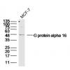
WB: 1:500
ICC: 1:50-1:200
IHC-P: 1:50-1:200
FC: 1:50-1:100
-
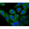
Fig1: ICC staining CaV2.3 in LOVO cells (green). The nuclear counter stain is DAPI (blue). Cells were fixed in paraformaldehyde, permeabilised with 0.25% Triton X100/PBS.
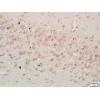
Fig2: Immunohistochemical analysis of paraffin-embedded rat brain tissue using anti-CaV2.3 antibody. Counter stained with hematoxylin.
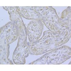
Fig3: Immunohistochemical analysis of paraffin-embedded human placenta tissue using anti-CaV2.3 antibody. Counter stained with hematoxylin.
Fig4: Immunohistochemical analysis of paraffin-embedded mouse testis tissue using anti-CaV2.3 antibody. Counter stained with hematoxylin.
Fig5: Flow cytometric analysis of 293T cells with CaV2.3 antibody at 1/100 dilution (fuchsia) compared with an unlabelled control (cells without incubation with primary antibody; yellow). Alexa Fluor 488-conjugated goat anti-rabbit IgG was used as the secondary antibody.
特别提示:本公司的所有产品仅可用于科研实验,严禁用于临床医疗及其他非科研用途!