Anti-CD34 antibody
-
概述
- 产品描述CD34 is a heavily glycosylated type I transmembrane molecule, that can be phoshorylated by a variety of kinases including Protein kinase C and Tyrosine kinases. CD34 antigen is expressed on small vessel endothelial cells and tumors of epithelial origin. CD34 is possibly an adhesion molecule with a putative role in early hematopoiesis by mediating the attachment of stem cells to the bone marrow extracellular matrix or directly to stromal cells. It could act as a scaffold for the attachment of lineage specific glycans, allowing stem cells to bind to lectins expressed by stromal cells or other marrow components. So CD34 is considered to be ideal marker for identifying and quantifying hematopoietic progenitor stem cells. With interaction with CrkL, CD34 becomes a substrate for PKC and CD34 surface expression is associated with activation of PKC.
- 产品名称Anti-CD34 antibody
- 分子量120 kDa
- 种属反应性Human,Mouse,Rat
- 验证应用WB, ICC, IHC-P, FC
- 抗体类型兔多抗
- 免疫原Synthetic peptide.
- 偶联Non-conjugated
-
性能
- 形态Liquid
- 浓度1 mg/mL.
- 存放说明Store at +4℃ after thawing. Aliquot store at -20℃ or -80℃. Avoid repeated freeze / thaw cycles.
- 存储缓冲液1*PBS (pH7.4), 0.2% BSA, 40% Glycerol. Preservative: 0.05% Sodium Azide.
- 亚型IgG
- 纯化方式Peptide affinity purified
- 亚细胞定位Cell membrane.
- 其它名称CD34 antibody
CD34 antigen antibody
CD34 molecule antibody
CD34_HUMAN antibody
Cluster designation 34 antibody
Hematopoietic progenitor cell antigen CD34 antibody
HPCA1 antibody
Mucosialin antibody
OTTHUMP00000034733 antibody
OTTHUMP00000034734 antibody
more
-
应用
WB: 1:1,000-1:2,000
ICC: 1:200
IHC-P: 1:2,000-1:3,000
FC: 1:50-1:100
-
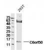
Fig1: Western blot analysis of CD34 on different cell lysates using anti-CD34antibody at 1/1000 dilution.
Positive control:
Lane 1: Human brain
Lane 2: Mouse brain
Lane 3: Mouse testis
Lane 4: Mouse thymus
Lane 5: Mouse spleen
Lane 6:Jurkat
Lane 7: TF-1
Lane 8: F9
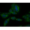
Fig2: ICC staining CD34 in hES cells (green). Cells were fixed in paraformaldehyde, permeabilised with 0.25% Triton X100/PBS.
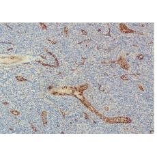
Fig3: Immunohistochemical analysis of paraffin-embedded human tonsil tissue using anti-CD34 antibody. Counter stained with hematoxylin.
Fig4: Immunohistochemical analysis of paraffin-embedded human liver carcinoma tissue using anti-CD34 antibody. Counter stained with hematoxylin.
Fig5: Flow cytometric analysis of Jurkat cells with CD34 antibody at 1/50 dilution (blue) compared with an unlabelled control (cells without incubation with primary antibody; red). Goat anti rabbit IgG (FITC) was used as the secondary antibody.
特别提示:本公司的所有产品仅可用于科研实验,严禁用于临床医疗及其他非科研用途!


