
![Anti-CD93 antibody [E10-A10]](images/202012/goods_img/92808_G_1608034521735.jpg)


-

专业包装 正品保证
-

快乐服务 售后无忧
-

会员特权 优惠不断
-

个人信息 严格保护
| 货号 | 规格 | 可用库存 | 销售价(RMB) | 您的折扣价(RMB) | 购买数量 |
|---|
| 熔点: | |
|---|---|
| 密度: | |
| 储存条件: | -20℃ |
Anti-CD93 antibody [E10-A10]
产品描述The CD93 antigen is a 652 amino acid cell-surface glycoprotein expressed by monocytes, neutrophils, platelets, microglia, and endothelial cells. CD93 was originally thought to be a putative receptor for the complement component C1q, a serum glycoprotein which plays an integral role in the activation of the classical pathway in response to immune complexes. As a result, in the literature the CD93 gene product has also been referred to as C1QR1 and C1qRp as well as MXRA4 (Matrix-remodeling-associated protein 4). Recent studies suggest that the CD93 antigen plays a role in intercellular adhesion and in clearance of apoptotic cells. CD93 is a heavily O-glycosylated, type I transmembrane protein consisting of an N-terminal domain with homology to C-type lectin domains, a tandem array of EGF-like domains, a single transmembrane domain and a short cytoplasmic tail.
产品名称Anti-CD93 antibody [E10-A10]
分子量69 kDa
种属反应性Human
验证应用WB,ICC,IHC-P,FC
抗体类型小鼠单抗
免疫原Recombinant protein
偶联Non-conjugated
形态Liquid
浓度2 mg/mL.
存放说明Store at +4℃ after thawing. Aliquot store at -20℃ or -80℃. Avoid repeated freeze / thaw cycles.
存储缓冲液1*TBS (pH7.4), 1%BSA, 40%Glycerol. Preservative: 0.05% Sodium Azide.
亚型IgG1
纯化方式Protein A purified.
亚细胞定位Membrane.
其它名称
C1q receptor 1 antibody
C1q/MBL/SPA receptor antibody
C1qR antibody
WB: 1:500-1:1,000
ICC: 1:50-1:200
IHC-P: 1:50-1:200
FC: 1:100-1:200

Fig1: Western blot analysis of CD93 on human CD93 recombinant protein using anti-CD93 antibody at 1/1,000 dilution.

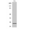
Fig2: Western blot analysis of CD93 on HEK293 (1) and CD93-hIgGFc transfected HEK293 (2) cell lysate using anti-CD93 antibody at 1/1,000 dilution.

Fig3: ICC staining CD93 (green) in Hela cells. The nuclear counter stain is DAPI (blue). Cells were fixed in paraformaldehyde, permeabilised with 0.25% Triton X100/PBS.

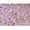
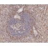
Fig4: Immunohistochemical analysis of paraffin-embedded human cervical cancer tissue using anti-CD93 antibody. Counter stained with hematoxylin.

Fig5: Flow cytometric analysis of HepG2 cells with CD93 antibody at 1/100 dilution (green) compared with an unlabelled control (cells without incubation with primary antibody; red).
特别提示:本公司的所有产品仅可用于科研实验,严禁用于临床医疗及其他非科研用途!


