Anti-Desmin antibody [3-F7]
-
概述
- 产品描述Desmin is one of the earliest protein markers for muscle tissue in embryogenesis as it is detected in the somites. Although it is present early in the development of muscle cells, it is only expressed at low levels, and increases as the cell nears terminal differentiation. Desmin is also important in muscle cell architecture and structure since it connects many components of the cytoplasm. Finally, desmin may be important in mitochondria function. Desmin-related myopathy (DRM or Desminopathy) is a subgroup of the myofibrillar myopathy diseases and is the result of a mutation in the gene that codes for desmin which prevents it from forming protein filaments, instead forming aggregates of desmin and other proteins throughout the cell. Recently, mutations were identified in patients suffered by an arrhythmogenic right ventricular cardiomyopathy (ARVC).
- 产品名称Anti-Desmin antibody [3-F7]
- 分子量53 kDa
- 种属反应性Human,Mouse
- 验证应用WB,ICC,IF,IHC-P,FC
- 抗体类型小鼠单抗
- 免疫原This antibody is produced by immunizing mice with recombinant protein of Desmin.
- 偶联Non-conjugated
-
性能
- 形态Liquid
- 浓度2 mg/mL.
- 存放说明Store at +4℃ after thawing. Aliquot store at -20℃. Avoid repeated freeze / thaw cycles.
- 存储缓冲液1*PBS (pH7.4), 0.2% BSA, 40% Glycerol. Preservative: 0.05% Sodium Azide.
- 亚型IgG2b
- 纯化方式Protein A purified.
- 亚细胞定位Cytopasm.
- 其它名称
- CMD1I antibody
- CSM1 antibody
- CSM2 antibody
more
-
应用
WB:1:2,000
ICC:1:200
IF:1:100
IHC-P:1:50-1:200
FC:1:50-1:100
-
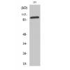
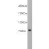
Fig1: Western blot analysis of Desmin on different lysates. Proteins were transferred to a PVDF membrane and blocked with 5% BSA in PBS for 1 hour at room temperature. The primary antibody was used at a 1:500 dilution in 5% BSA at room temperature for 2 hours. Goat Anti-Mouse IgG - HRP Secondary Antibody (HA1006) at 1:5,000 dilution was used for 1 hour at room temperature.
Positive control:
Lane 1: Human skeletal muscle tissue lysate, untreated
Lane 2: Human heart tissue lysate, untreated
Fig2: ICC staining Desmin in D3 cells (green). Formalin fixed cells were permeabilized with 0.1% Triton X-100 in TBS for 10 minutes at room temperature and blocked with 1% Blocker BSA for 15 minutes at room temperature. Cells were probed with Desmin monoclonal antibody at a dilution of 1:100 for 1 hour at room temperature, washed with PBS. Alexa Fluorc™ 488 Goat anti-Mouse IgG was used as the secondary antibody at 1/100 dilution.
Fig3: ICC staining Desmin in Hela cells (green). Formalin fixed cells were permeabilized with 0.1% Triton X-100 in TBS for 10 minutes at room temperature and blocked with 1% Blocker BSA for 15 minutes at room temperature. Cells were probed with Desmin monoclonal antibody at a dilution of 1:100 for 1 hour at room temperature, washed with PBS. Alexa Fluorc™ 488 Goat anti-Mouse IgG was used as the secondary antibody at 1/100 dilution. The nuclear counter stain is DAPI (blue).
Fig4: ICC staining Desmin in HepG2 cells (green). Formalin fixed cells were permeabilized with 0.1% Triton X-100 in TBS for 10 minutes at room temperature and blocked with 1% Blocker BSA for 15 minutes at room temperature. Cells were probed with Desmin monoclonal antibody at a dilution of 1:100 for 1 hour at room temperature, washed with PBS. Alexa Fluorc™ 488 Goat anti-Mouse IgG was used as the secondary antibody at 1/100 dilution.
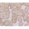
Fig5: Immunohistochemical analysis of paraffin-embedded human cervix tissue using anti-Desmin antibody. The section was pre-treated using heat mediated antigen retrieval with Tris-EDTA buffer (pH 8.0-8.4) for 20 minutes.The tissues were blocked in 5% BSA for 30 minutes at room temperature, washed with ddH2O and PBS, and then probed with the antibody at 1/100 dilution, for 30 minutes at room temperature and detected using an HRP conjugated compact polymer system. DAB was used as the chrogen. Counter stained with hematoxylin and mounted with DPX.
Fig6: Immunohistochemical analysis of paraffin-embedded human stomach cancer tissue using anti-Desmin antibody. The section was pre-treated using heat mediated antigen retrieval with Tris-EDTA buffer (pH 8.0-8.4) for 20 minutes.The tissues were blocked in 5% BSA for 30 minutes at room temperature, washed with ddH2O and PBS, and then probed with the antibodyat 1/100 dilution, for 30 minutes at room temperature and detected using an HRP conjugated compact polymer system. DAB was used as the chrogen. Counter stained with hematoxylin and mounted with DPX.
Fig7: Immunohistochemical analysis of paraffin-embedded human uterus tissue using anti-Desmin antibody. The section was pre-treated using heat mediated antigen retrieval with Tris-EDTA buffer (pH 8.0-8.4) for 20 minutes.The tissues were blocked in 5% BSA
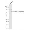
Fig8: Flow cytometric analysis of Desmin was done on Hela cells. The cells were fixed, permeabilized and stained with Desmin antibody at 1/100 dilution (blue) compared with an unlabelled control (cells without incubation with primary antibody; red). After
特别提示:本公司的所有产品仅可用于科研实验,严禁用于临床医疗及其他非科研用途!


![Anti-Desmin antibody [3-F7]](images/202012/goods_img/92413_G_1607327981791.jpg)


