Anti-MIB1 antibody [C9-A9]
-
概述
- 产品描述The LIN-12/notch family of transmembrane receptors is believed to play a central role in development by regulating cell fate decisions. MIB1 (E3 ubiquitin-protein ligase MIB1), also known as Mind bomb homolog 1 and DAPK-interacting protein 1, is a 1006 amino acid E3 ubiquitin ligase that activates the Notch ligand, Delta. MIB1 ubiquinates Delta by binding to its intracellular domain, leading to the endocytosis and eventual degradation of the Delta receptor, which, paradoxically, results in the up-regulation of receptor activity and enhances Notch signaling. MIB1 also interacts with DAPK, a protein that plays an important role in the regulation of apoptosis. Ubiquination of DAPK leads to inhibition of caspase-dependent apoptosis, therefore it is likely that overexpression of MIB1 can lead to tumor growth. Although it seems to be widely expressed at low levels, MIB1 is expressed at highest concentrations in the CNS and ovary. Both DAPK and MIB1 are overexpressed in epileptic brain tissue, suggesting that they probably cooperate as regulators of neuronal death in epilepsy.
- 产品名称Anti-MIB1 antibody [C9-A9]
- 分子量110 kDa
- 种属反应性Human,Monkey
- 验证应用WB,ICC,FC
- 抗体类型小鼠单抗
- 免疫原Recombinant protein
- 偶联Non-conjugated
-
性能
- 形态Liquid
- 浓度2 mg/mL.
- 存放说明Store at +4℃ after thawing. Aliquot store at -20℃ or -80℃. Avoid repeated freeze / thaw cycles.
- 存储缓冲液1*TBS (pH7.4), 1%BSA, 40%Glycerol. Preservative: 0.05% Sodium Azide.
- 亚型IgG1
- 纯化方式Protein A purified.
- 亚细胞定位Cytoplasm. Cytoskeleton. Cell membrane
- 其它名称
- DAPK-interacting protein 1 antibody
- Dip 1 antibody
- DIP-1 antibody
more
-
应用
WB: 1:500-1:2,000
ICC: 1:100-1:500
FC: 1:100-1:200
-
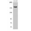
Fig1: Western blot analysis of MIB1 on human MIB1 recombinant protein using anti-MIB1 antibody at 1/1,000 dilution.
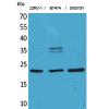
Fig2: Western blot analysis of MIB1 on HEK293 (1) and MIB1-hIgGFc transfected HEK293 (2) cell lysate using anti-MIB1 antibody at 1/1,000 dilution.
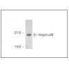
Fig3: Western blot analysis of MIB1 on Hela (1) and COS7 (2) cell lysate using anti-MIB1 antibody at 1/1,000 dilution.
Fig4: ICC staining MIB1 (green) and Actin filaments (red) in Hela cells. The nuclear counter stain is DAPI (blue). Cells were fixed in paraformaldehyde, permeabilised with 0.25% Triton X100/PBS.
Fig5: Flow cytometric analysis of Hela cells with MIB1 antibody at 1/100 dilution (green) compared with an unlabelled control (cells without incubation with primary antibody; red).
特别提示:本公司的所有产品仅可用于科研实验,严禁用于临床医疗及其他非科研用途!


![Anti-MIB1 antibody [C9-A9]](images/202012/goods_img/92149_G_1606987195101.jpg)


