Anti-ESR1 antibody [E8-A6]
-
概述
- 产品描述Estrogen receptors (ER) are members of the steroid/thyroid hormone receptor superfamily of ligand-activated transcription factors. Estrogen receptors, including ERα and ERβ, contain DNA binding and ligand binding domains and are critically involved in regulating the normal function of reproductive tissues. They are located in the nucleus, though some estrogen receptors associate with the cell surface membrane and can be rapidly activated by exposure of cells to estrogen. ERα and ERβ have been shown to be differentially activated by various ligands. Receptor-ligand interactions trigger a cascade of events, including dissociation from heat shock proteins, receptor dimerization, phosphorylation and the association of the hormone activated receptor with specific regulatory elements in target genes. Evidence suggests that ERα and ERβ may be regulated by distinct mechanisms even though they share many functional characteristics.
- 产品名称Anti-ESR1 antibody [E8-A6]
- 分子量66 kDa
- 种属反应性Human,Rat
- 验证应用WB,ICC,IHC-P,FC
- 抗体类型小鼠单抗
- 免疫原Recombinant protein
- 偶联Non-conjugated
-
性能
- 形态Liquid
- 浓度2 mg/mL.
- 存放说明Store at +4℃ after thawing. Aliquot store at -20℃ or -80℃. Avoid repeated freeze / thaw cycles.
- 存储缓冲液1*TBS (pH7.4), 1%BSA, 40%Glycerol. Preservative: 0.05% Sodium Azide.
- 亚型IgG1
- 纯化方式Protein A purified.
- 亚细胞定位Nucleus, cytoplasm.
- 其它名称
- DKFZp686N23123 antibody
- ER alpha antibody
- ER antibody
more
-
应用
WB: 1:500-1:1,000
ICC: 1:50-1:200
IHC-P: 1:50-1:200
FC: 1:50-1:100
-
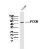
Fig1: Western blot analysis of ESR1 on human ESR1 recombinant protein using anti-ESR1 antibody at 1/1,000 dilution.
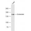
Fig2: Western blot analysis of ESR1 on HEK293 (1) and ESR1-hIgGFc transfected HEK293 (2) cell lysate using anti-ESR1 antibody at 1/1,000 dilution.
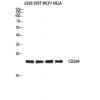
Fig3: Western blot analysis of ESR1 on different cell lysate using anti-ESR1 antibody at 1/1,000 dilution.
Positive control: Line1: MOLT4 Line2: Raji Line3: MCF-7 Line4: T47D Line5: SK-Br-3 Line6: Hela Line7: C6
Fig4: ICC staining ESR1 (green) and Actin filaments (red) in Hela cells. Cells were fixed in paraformaldehyde, permeabilised with 0.25% Triton X100/PBS.
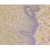
Fig5: Immunohistochemical analysis of paraffin-embedded human breast cancer tissue using anti-ESR1 antibody. Counter stained with hematoxylin.
Fig6: Immunohistochemical analysis of paraffin-embedded human esophageal cancer tissue using anti-ESR1 antibody. Counter stained with hematoxylin.
Fig7: Flow cytometric analysis of Hela cells with ESR1 antibody at 1/100 dilution (green) compared with an unlabelled control (cells without incubation with primary antibody; red).
特别提示:本公司的所有产品仅可用于科研实验,严禁用于临床医疗及其他非科研用途!

![Anti-ESR1 antibody [E8-A6]](images/202012/goods_img/92113_G_1606983832711.jpg)