Anti-MEF2C antibody [G2-H2]
-
概述
- 产品描述The myocyte enhancer factor-2 (MEF-2) family of transcription factors associate with co-repessors or co-activators to regulate development and function of T cells, neuronal cells, and muscle cells. Four family members arise from alternatively spliced transcripts, termed MEF-2A, -2B, -2C, and -2D. These members bind as homo- and heterodimers to the MEF-2 site in the promoter region of affected genes. Differential regulation in the expression of the four transcripts implies functional distinction for each duing embryogenesis and development. The process of differentiation from mesodermal precursor cells to myoblasts has led to the discovery of a variety of tissue-specific factors that regulate muscle gene expression. The myogenic basic helix-loop-helix proteins, including MyoD, myogenin, Myf-5, and MRF4, are one class of identified factors. A second family of DNA binding regulatory proteins is the myocyte-specific enhancer factor-2 (MEF-2) family. Each of these proteins binds to the MEF-2 target DNA sequence present in the regulatory regions of many muscle-specific genes.
- 产品名称Anti-MEF2C antibody [G2-H2]
- 分子量51 kDa
- 种属反应性Human,Mouse
- 验证应用WB,IHC-P,FC
- 抗体类型小鼠单抗
- 免疫原Recombinant protein
- 偶联Non-conjugated
-
性能
- 形态Liquid
- 浓度2 mg/mL.
- 存放说明Store at +4℃ after thawing. Aliquot store at -20℃ or -80℃. Avoid repeated freeze / thaw cycles.
- 存储缓冲液1*TBS (pH7.4), 1%BSA, Preservative: 0.05% Sodium Azide.
- 亚型IgG1
- 纯化方式Protein A purified.
- 亚细胞定位Nucleus.
- 其它名称
- C5DELq14.3 antibody
- DEL5q14.3 antibody
- MADS box transcription enhancer factor 2 polypeptide C (myocyte enhancer factor 2C) antibody
more
-
应用
WB: 1:500
IHC-P: 1:100-1:200
FC: 1:100-1:200
-
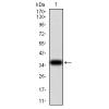
Fig1: Western blot analysis of MEF2C on human MEF2C recombinant protein using anti-MEF2C antibody at 1/1,000 dilution.
Fig2: Western blot analysis of MEF2C on HEK293 (1) and MEF2C-hIgGFc transfected HEK293 (2) cell lysate using anti-MEF2C antibody at 1/1,000 dilution.
Fig3: Western blot analysis of MEF2C on NIH3T3 (1) and 3T3-L1 (2) cell lysate using anti-MEF2C antibody at 1/1,000 dilution.
Fig4: Immunohistochemical analysis of paraffin-embedded human colon cancer tissue using anti-MEF2C antibody. Counter stained with hematoxylin.
Fig5: Immunohistochemical analysis of paraffin-embedded human esophageal cancer tissue using anti-MEF2C antibody. Counter stained with hematoxylin.
Fig6: Flow cytometric analysis of Hela cells with MEF2C antibody at 1/100 dilution (green) compared with an unlabelled control (cells without incubation with primary antibody; purple).
特别提示:本公司的所有产品仅可用于科研实验,严禁用于临床医疗及其他非科研用途!

![Anti-MEF2C antibody [G2-H2]](images/202012/goods_img/92102_G_1606982954468.jpg)