Anti-HSP47 antibody
-
概述
- 产品描述Heat shock protein 47, also known as SERPINH1 is a serpin which serves as a human chaperone protein for collagen. This protein is a member of the serpin superfamily of serine proteinase inhibitors. Its expression is induced by heat shock. HSP47 is expressed by cells present in Endoplasmic Reticulum. These cells synthesize and secrete type I and type II collagen. The protein localizes to the endoplasmic reticulum lumen and binds collagen; thus it is thought to be a molecular chaperone involved in the maturation of collagen molecules. Autoantibodies to this protein have been found in patients with rheumatoid arthritis. Heat shock protein 47 has been shown to interact with collagens I, II, III, IV and V. In the ER, HSP47 interacts with and stabilizes correctly-folded procollagen. Nucleotide polymorphisms may be associated with preterm birth and Osteogenesis Imperfecta type X. Serpin-H1 is up-regulated in several cancers including squamous cell carcinoma, breast and prostate carcinomas. Expression in tumors drives malignant growth and invasion by enhancing deposition of extracellular matrix proteins.
- 产品名称Anti-HSP47 antibody
- 分子量46 kDa.
- 种属反应性Human, Mouse,Rat
- 验证应用WB,IHC-P
- 抗体类型兔多抗
- 免疫原Recombinant protein within human HSP47 aa 82-318.
- 偶联Non-conjugated
-
性能
- 形态Liquid
- 浓度1 mg/mL.
- 存放说明Store at +4℃ after thawing. Aliquot store at -20℃ or -80℃. Avoid repeated freeze / thaw cycles.
- 存储缓冲液1*PBS (pH7.4), 0.2% BSA, 40% Glycerol. Preservative: 0.05% Sodium Azide.
- 亚型IgG
- 纯化方式Protein affinity purified.
- 亚细胞定位Endoplasmic reticulum.
- 其它名称
- 47 kDa heat shock protein antibody
- 47 kDa heat shock protein precursor antibody
- Arsenic transactivated protein 3 antibody
more
-
应用
WB: 1:500-1:2,000
IHC-P: 1:100-1:500
-
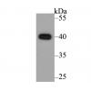
Fig1: Western blot analysis of HSP47 on different lysates. Proteins were transferred to a PVDF membrane and blocked with 5% BSA in PBS for 1 hour at room temperature. The primary antibody was used in 5% BSA at room temperature for 2 hours. Goat Anti-Rabbit IgG - HRP Secondary Antibody (HA1001) at 1:5,000 dilution was used for 1 hour at room temperature.
Positive control:
Lane 1: Rat placenta tissue lysate
Lane 2: Siha cell lysate
Lane 3: NIH/3T3 cell lysate
Fig2: Immunohistochemical analysis of paraffin-embedded human placenta tissue using anti-HSP47 antibody. The section was pre-treated using heat mediated antigen retrieval with sodium citrate buffer (pH 6.0) for 20 minutes. The tissues were blocked in 5% BSA for 30 minutes at room temperature, washed with ddH2O and PBS, and then probed with the primary antibody ( for 30 minutes at room temperature. The detection was performed using an HRP conjugated compact polymer system. DAB was used as the chromogen. Tissues were counterstained with hematoxylin and mounted with DPX.
特别提示:本公司的所有产品仅可用于科研实验,严禁用于临床医疗及其他非科研用途!