Anti-HIF-1 alpha antibody
-
概述
- 产品描述Cell growth and viability is compromised by oxygen deprivation (hypoxia). Hypoxia-inducible factors, including HIF-1α, HIF-1β (also designated Arnt 1), EPAS-1 (also designated HIF-2α) and HIF-3α, induce glycolysis, erythropoiesis and angiogenesis in order to restore oxygen homeostasis. Hypoxia-inducible factors are members of the Per-Arnt-Sim (PAS) domain transcription factor family. In response to hypoxia, HIF-1α is upregulated and forms a heterodimer with Arnt 1 to form the HIF-1 complex. The HIF-1 complex recognizes and binds to the hypoxia responsive element (HRE) of hypoxia-inducible genes, thereby activating transcription. Hypoxia-inducible expression of some genes such as Glut-1, p53, p21 or Bcl-2, is HIF-1α dependent, whereas expression of others, such as p27, GADD 153 or HO-1, is HIF-1α independent. EPAS-1 and HIF-3α have also been shown to form heterodimeric complexes with Arnt 1 in response to hypoxia.
- 产品名称Anti-HIF-1 alpha antibody
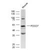
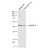
- 分子量92 kDa
- 种属反应性Human,Mouse,Rat
- 验证应用WB,ICC,IHC-P,FC
- 抗体类型兔多抗
- 免疫原peptide
- 偶联Non-conjugated
-
性能
- 形态Liquid
- 浓度1 mg/mL.
- 存放说明Store at +4℃ after thawing. Aliquot store at -20℃ or -80℃. Avoid repeated freeze / thaw cycles.
- 存储缓冲液1*PBS (pH7.4), 0.2% BSA, 40% Glycerol. Preservative: 0.05% Sodium Azide.
- 亚型IgG
- 纯化方式Peptide affinity purified
- 亚细胞定位Cytoplasm, Nucleus, Nucleus speckle
- 其它名称
- ARNT interacting protein antibody
- ARNT-interacting protein antibody
- Basic helix loop helix PAS protein MOP1 antibody
more
-
应用
ICC: 1:50-1:200
IHC-P: 1:50-1:200
FC: 1:50-1:100
WB: 1:500
-
Fig1: Immunocytochemical staining of Hela cells using anti-HIF-1 alpha rabbit polyclonal antibody. .
Fig2: Immunocytochemical staining of PANC-1 cells using anti-HIF-1 alpha rabbit polyclonal antibody. .
Fig3: Immunocytochemical staining of SW480 cells using anti-HIF-1 alpha rabbit polyclonal antibody. .
Fig4: Immunohistochemical analysis of paraffin- embedded human tonsil tissue using anti-HIF-1 alpha rabbit polyclonal antibody. .
Fig5: Immunohistochemical analysis of paraffin- embedded human colon carcinoma tissue using anti-HIF-1 alpha rabbit polyclonal antibody. .
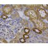
Fig6: Immunohistochemical analysis of paraffin- embedded mouse colon tissue using anti-HIF-1 alpha rabbit polyclonal antibody. .
Fig7: Immunohistochemical analysis of paraffin- embedded mouse small intestine tissue using anti-HIF-1 alpha rabbit polyclonal antibody. .
Fig8: Flow cytometric analysis of Hela cells with HIF-1 alpha antibody at 1/100 dilution (blue) compared with an unlabelled control (cells without incubation with primary antibody; red). Alexa Fluor 488-conjugated Goat anti rabbit IgG was used as the seco
特别提示:本公司的所有产品仅可用于科研实验,严禁用于临床医疗及其他非科研用途!