Anti-PHD2 antibody
-
概述
- 产品描述Prolyl hydroxylase domain proteins HIF PHD1, HIF PHD2 and HIF PHD3 (known as PHD1, PHD2 and PHD3 in rodents, respectively) can hydroxylate HIF-α subunits. Hypoxia-inducible factor (HIF) is a transcriptional regulator important in several aspects of oxygen homeostasis. The prolyl hydroxylases catalyze the posttranslational formation of 4-hydroxyproline in HIF-α proteins. HIF PHD1, which is widely expressed, with highest levels of expression in testis, functions as a cellular oxygen sensor and is important in cell growth regulation. HIF PHD1 can localize to the nucleus or the cytoplasm and is also detected in hormone responsive tissues, such as normal and cancerous mammary, ovarian and prostate epithelium. HIF PHD1 is encoded by EGLN2, which maps to chromosome 19q13.3. HIF PHD2 is regarded as the main cellular oxygen sensor, as RNA interference against HIF PHD2, but not HIF PHD1 or HIF PHD3, is enough to stabilize HIF-1α in normoxia. HIF PHD2, a direct HIF target gene, is expressed mainly in skeletal muscle, heart, kidney and brain.
- 产品名称Anti-PHD2 antibody
- 分子量46kDa
- 种属反应性Human,Mouse,Rat
- 验证应用WB,ICC,IHC-P,FC
- 抗体类型兔多抗
- 免疫原peptide
- 偶联Non-conjugated
-
性能
- 形态Liquid
- 浓度1 mg/mL.
- 存放说明Store at +4℃ after thawing. Aliquot store at -20℃ or -80℃. Avoid repeated freeze / thaw cycles.
- 存储缓冲液1*PBS (pH7.4), 0.2% BSA, 40% Glycerol. Preservative: 0.05% Sodium Azide.
- 亚型IgG
- 纯化方式Peptide affinity purified
- 亚细胞定位Cytoplasm, Nucleus
- 其它名称
- C1ORF12 antibody
- Chromosome 1 Open Reading Frame 12 antibody
- DKFZp761F179 antibody
more
-
应用
WB: 1:1,000
ICC: 1:50-1:200
IHC-P: 1:50-1:200
FC: 1:10-1:100
-
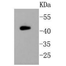
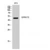
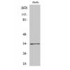
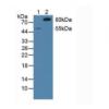
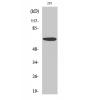
Fig1: Western blot analysis on mouse brain lysates using anti-PHD2 rabbit polyclonal antibody.
Fig2: Immunocytochemical staining of PC-12 cells using anti-PHD2 rabbit polyclonal antibody.
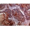
Fig3: Immunohistochemical analysis of paraffin- embedded human kidney tissue using anti-PHD2 rabbit polyclonal antibody.
Fig4: Immunohistochemical analysis of paraffin- embedded human pancreas tissue using anti-PHD2 rabbit polyclonal antibody.
Fig5: Immunohistochemical analysis of paraffin- embedded mouse brain tissue using anti-PHD2 rabbit polyclonal antibody.
Fig6: Immunohistochemical analysis of paraffin- embedded mouse kidney tissue using anti-PHD2 rabbit polyclonal antibody.
Fig7: Flow cytometric analysis of SH-SY-5Y cells with PHD2 antibody at 1/50 dilution (blue) compared with an unlabelled control (cells without incubation with primary antibody; red). Alexa Fluor 488-conjugated Goat anti rabbit IgG was used as the secondar
特别提示:本公司的所有产品仅可用于科研实验,严禁用于临床医疗及其他非科研用途!


