Anti-PDCD6 antibody
-
概述
- 产品描述This gene encodes a calcium-binding protein belonging to the penta-EF-hand protein family. Calcium binding is important for homodimerization and for conformational changes required for binding to other protein partners. This gene product participates in T cell receptor-, Fas-, and glucocorticoid-induced programmed cell death. In mice deficient for this gene product, however, apoptosis was not blocked suggesting this gene product is functionally redundant. Alternatively spliced transcript variants encoding multiple isoforms have been observed for this gene, and a pseudogene of this gene is also located on the short arm of chromosome 5.
- 产品名称Anti-PDCD6 antibody
- 分子量22 kDa
- 种属反应性Human,Rat
- 验证应用WB,IHC-P
- 抗体类型兔多抗
- 免疫原Recombinant protein within human PDCD6 aa 1-191.
- 偶联Non-conjugated
-
性能
- 形态Liquid
- 浓度1 mg/mL.
- 存放说明Store at +4℃ after thawing. Aliquot store at -20℃. Avoid repeated freeze / thaw cycles.
- 存储缓冲液1*TBS (pH7.4), 0.2% BSA, 50% Glycerol. Preservative: 0.05% Sodium Azide.
- 亚型IgG
- 纯化方式Protein affinity purified.
- 亚细胞定位Endosome, Endoplasmic reticulum membrane, Nucleus, COPII-coated vesicle membrane, Cytoplasm.
- 其它名称
- AIP1 antibody
- ALG 2 antibody
- ALG-2-interacting protein 1 antibody
more
-
应用
WB: 1:500-1:2,000
IHC-P: 1:100-1:500
-
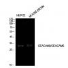
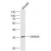
Fig1: Western blot analysis of PDCD6 on different lysates. Proteins were transferred to a PVDF membrane and blocked with 5% BSA in PBS for 1 hour at room temperature. The primary antibody ( was used in 5% BSA at room temperature for 2 hours. Goat Anti-Rabbit IgG - HRP Secondary Antibody (HA1001) at 1:5,000 dilution was used for 1 hour at room temperature.
Positive control:
Lane 1: SK-Br-3 cell lysate
Lane 2: HepG2 cell lysate
Lane 1: Rat ovary tissue lysate
Fig2: Immunohistochemical analysis of paraffin-embedded human breast carcinoma tissue using anti-PDCD6 antibody. The section was pre-treated using heat mediated antigen retrieval with sodium citrate buffer (pH 6.0) for 20 minutes. The tissues were blocked in 5% BSA for 30 minutes at room temperature, washed with ddH2O and PBS, and then probed with the primary antibody for 30 minutes at room temperature. The detection was performed using an HRP conjugated compact polymer system. DAB was used as the chromogen. Tissues were counterstained with hematoxylin and mounted with DPX.
Fig3: Immunohistochemical analysis of paraffin-embedded human placenta tissue using anti-PDCD6 antibody. The section was pre-treated using heat mediated antigen retrieval with sodium citrate buffer (pH 6.0) for 20 minutes. The tissues were blocked in 5% BSA for 30 minutes at room temperature, washed with ddH2O and PBS, and then probed with the primary antibodyfor 30 minutes at room temperature. The detection was performed using an HRP conjugated compact polymer system. DAB was used as the chromogen. Tissues were counterstained with hematoxylin and mounted with DPX.
Fig4: Immunohistochemical analysis of paraffin-embedded human stomach carcinoma tissue using anti-PDCD6 antibody. The section was pre-treated using heat mediated antigen retrieval with sodium citrate buffer (pH 6.0) for 20 minutes. The tissues were blocked in 5% BSA for 30 minutes at room temperature, washed with ddH2O and PBS, and then probed with the primary antibody for 30 minutes at room temperature. The detection was performed using an HRP conjugated compact polymer system. DAB was used as the chromogen. Tissues were counterstained with hematoxylin and mounted with DPX.
特别提示:本公司的所有产品仅可用于科研实验,严禁用于临床医疗及其他非科研用途!