Anti-ATP8A1 antibody
-
概述
- 产品描述Catalytic component of a P4-ATPase flippase complex which catalyzes the hydrolysis of ATP coupled to the transport of aminophospholipids from the outer to the inner leaflet of various membranes and ensures the maintenance of asymmetric distribution of phospholipids. Phospholipid translocation seems also to be implicated in vesicle formation and in uptake of lipid signaling molecules. In vitro, its ATPase activity is selectively and stereospecifically stimulated by phosphatidylserine (PS). The flippase complex ATP8A1:TMEM30A seems to play a role in regulation of cell migration probably involving flippase-mediated translocation of phosphatidylethanolamine (PE) at the plasma membrane. Acts as aminophospholipid translocase at the plasma membrane in neuronal cells.
- 产品名称Anti-ATP8A1 antibody
- 分子量131 kDa
- 种属反应性Human,Rat
- 验证应用WB,FC
- 抗体类型兔多抗
- 免疫原Recombinant protein within human ATP8A1 aa 400-700.
- 偶联Non-conjugated
-
性能
- 形态Liquid
- 浓度1 mg/ml.
- 存放说明Store at +4℃ after thawing. Aliquot store at -20℃. Avoid repeated freeze / thaw cycles.
- 存储缓冲液1*PBS (pH7.4), 0.2% BSA, 50% Glycerol. Preservative: 0.05% Sodium Azide.
- 亚型IgG
- 纯化方式Protein affinity purified.
- 亚细胞定位Cell membrane, Cytoplasmic vesicle, Endoplasmic reticulum, Golgi apparatus, Membrane.
- 其它名称
- Phospholipid-transporting ATPase IA antibody
- ATPase class I type 8A member 1 antibody
- Chromaffin granule ATPase II antibody
more
-
应用
WB: 1:500-1:1,000
FC: 1:50-1:100
-
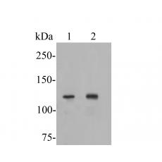
Fig1: Western blot analysis of ATP8A1 on different lysates. Proteins were transferred to a PVDF membrane and blocked with 5% BSA in PBS for 1 hour at room temperature. The primary antibody was used in 5% BSA at room temperature for 2 hours. Goat Anti-Rabbit IgG - HRP Secondary Antibody (HA1001) at 1:5,000 dilution was used for 1 hour at room temperature.
Positive control:
Lane 1: MCF-7 cell lysate
Lane 2: Rat brain tissue lysate
Fig2: Flow cytometric analysis of ATP8A1 was done on MCF-7 cells. The cells were fixed, permeabilized and stained with the primary antibody (yellow). After incubation of the primary antibody at room temperature for an hour, the cells were stained with a Alexa Fluor 488-conjugated goat anti-rabbit IgG Secondary antibody at 1/500 dilution for 30 minutes.Unlabelled sample was used as a control (cells without incubation with primary antibody; purple).
特别提示:本公司的所有产品仅可用于科研实验,严禁用于临床医疗及其他非科研用途!