Anti-MLCK antibody
-
概述
- 产品描述The Ca2+/calmodulin-dependent protein kinases (CaM kinases) are a structurally related subfamily of serine/threonine kinases that includes CaMKI, CaMKII, CaMKIV and myosin light chain kinases (MYLK, also designated MLCK). The MYLK kinases phosphorylate myosin regulatory light chains to catalyze myosin interaction with actin filaments resulting in contractile activity. Non-muscle, smooth muscle and skeletal/cardiac muscle MYLK isoforms exist. The MYLK gene (also designated MYLK1) encodes both smooth muscle and non-muscle isoforms as well as telokin, a small C-terminal isoform expressed only in smooth muscle with the capacity to stabilize unphosphorylated myosin filaments. Multiple transcript variants are described for the MYLK gene. Smooth-muscle and non-muscle MYLK isoforms are expressed in a wide variety of adult and fetal tissues. The skeletal/cardiac muscle isoforms of MYLK are encoded by a separate gene, MYLK2 (also designated skMLCK). MYLK appears to be a target for PAKs (p21-activated kinases). PAK1 interaction with MYLK results in a decrease in MYLK activity and myosin light chain phosphorylation.
- 产品名称Anti-MLCK antibody
- 种属反应性Human,Mouse
- 验证应用ICC,IHC-P,FC
- 抗体类型兔多抗
- 免疫原peptide
- 偶联Non-conjugated
-
性能
- 形态Liquid
- 浓度1 mg/mL.
- 存放说明Store at +4℃ after thawing. Aliquot store at -20℃ or -80℃. Avoid repeated freeze / thaw cycles.
- 存储缓冲液1*PBS (pH7.4), 0.2% BSA, 40% Glycerol. Preservative: 0.05% Sodium Azide.
- 亚型IgG
- 纯化方式Peptide affinity purified
- 亚细胞定位Cytoplasm, Cleavage furrow, Cell projection
- 其它名称
- deglutamylated form antibody
- DKFZp686I10125 antibody
- EC 2.7.11.18 antibody
more
-
应用
ICC: 1:50-1:200
ICC: 1:50-1:200
FC: 1:50-1:100
-
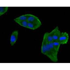
Fig1: Immunocytochemical staining of Hela cells using anti-MLCK rabbit polyclonal antibody.
Fig2: Immunocytochemical staining of NIH/3T3 cells using anti-MLCK rabbit polyclonal antibody.
Fig3: Immunocytochemical staining of SHG-44 cells using anti-MLCK rabbit polyclonal antibody.
Fig4: Immunohistochemical analysis of paraffin- embedded mouse smooth muscle tissue using anti-MLCK rabbit polyclonal antibody.
Fig5: Flow cytometric analysis of N2A cells with MLCK antibody at 1/50 dilution (blue) compared with an unlabelled control (cells without incubation with primary antibody; red). Alexa Fluor 488-conjugated Goat anti rabbit IgG was used as the secondary antibody.
特别提示:本公司的所有产品仅可用于科研实验,严禁用于临床医疗及其他非科研用途!