Anti-nNOS antibody
-
概述
- 产品描述Nitric oxide (NO) has a broad range of biological activities and has been implicated in signaling pathways in phylogenetically diverse species. Nitric oxide synthases (NOSs), the enzymes responsible for synthesis of NO, contain an N-terminal oxygenase domain and a C-terminal reductase domain. NOS activity requires homodimerization as well as three cosubstrates (L-arginine, NADPH and O2) and five cofactors or prosthetic groups (FAD, FMN, calmodulin, tetrahydrobiopterin and heme). Several distinct NOS isoforms have been described and been shown to represent the products of three distinct genes. These include two constitutive Ca2+/CaM-dependent forms of NOS, including NOS1 (also designated ncNOS) whose activity was first identified in neurons, and NOS3 (also designated ecNOS), first identified in endothelial cells. The inducible form of NOS, NOS2 (also designated iNOS), is Ca2+-independent and is expressed in a broad range of cell types.
- 产品名称Anti-nNOS antibody
- 分子量160kDa
-
种属反应性Human,Mouse,Rat
-
验证应用WB,ICC,IHC-P,FC
-
抗体类型兔多抗
-
免疫原peptide
-
偶联Non-conjugated
-
性能
-
形态Liquid
-
浓度1 mg/mL.
-
存放说明Store at +4℃ after thawing. Aliquot store at -20℃ or -80℃. Avoid repeated freeze / thaw cycles.
-
存储缓冲液1*PBS (pH7.4), 0.2% BSA, 40% Glycerol. Preservative: 0.05% Sodium Azide.
-
亚型IgG
-
纯化方式Peptide affinity purified
-
亚细胞定位Cell membrane, Cell projection
-
-
其它名称
more
-
应用
WB: 1:1,000
ICC: 1:50-1:200
IHC-P: 1:50-1:200
FC: 1:50-1:100
-
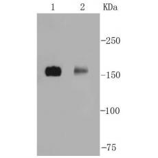
Fig1: Western blot analysis on SHG-44(1) and mouse brain(2) lysates using anti-nNOS rabbit polyclonal antibody.
Fig2: Immunocytochemical staining of SK-BR-3 cells using anti-nNOS rabbit polyclonal antibody.
Fig3: Immunocytochemical staining of HepG2 cells using anti-nNOS rabbit polyclonal antibody.
Fig4: Immunohistochemical analysis of paraffin- embedded human lung tissue using anti-nNOS rabbit polyclonal antibody.
Fig5: Immunohistochemical analysis of paraffin- embedded human kidney tissue using anti-nNOS rabbit polyclonal antibody.
Fig6: Immunohistochemical analysis of paraffin- embedded mouse lung tissue using anti-nNOS rabbit polyclonal antibody.
Fig7: Immunohistochemical analysis of paraffin- embedded mouse testis tissue using anti-nNOS rabbit polyclonal antibody.
Fig8: Immunohistochemical analysis of paraffin- embedded mouse brain tissue using anti-nNOS rabbit polyclonal antibody.
Fig9: Immunohistochemical analysis of paraffin- embedded mouse kidney tissue using anti-nNOS rabbit polyclonal antibody.
Fig10: Flow cytometric analysis of N2A cells with nNOS antibody at 1/50 dilution (blue) compared with an unlabelled control (cells without incubation with primary antibody; red). Alexa Fluor 488-conjugated Goat anti rabbit IgG was used as the secondary an
特别提示:本公司的所有产品仅可用于科研实验,严禁用于临床医疗及其他非科研用途!