


-

专业包装 正品保证
-

快乐服务 售后无忧
-

会员特权 优惠不断
-

个人信息 严格保护
| 别名: | p70 S6 Kinase beta 1 | ||
|---|---|---|---|
| 适用物种: | Human, Mouse, Rat | ||
| 验证应用: | WB, ICC, IHC-P, FC | ||
| 种属: | 兔多抗 | ||
| 储存条件: | -20℃ | ||
|
| 货号 | 规格 | 可用库存 | 销售价(RMB) | 您的折扣价(RMB) | 购买数量 |
|---|
| 熔点: | |
|---|---|
| 密度: | |
| 储存条件: | -20℃ |
Anti-p70 S6 Kinase beta 1 antibody
产品描述Serine/threonine-protein kinase that acts downstream of mTOR signaling in response to growth factors and nutrients to promote cell proliferation, cell growth and cell cycle progression. Regulates protein synthesis through phosphorylation of EIF4B, RPS6 and EEF2K, and contributes to cell survival by repressing the pro-apoptotic function of BAD. Under conditions of nutrient depletion, the inactive form associates with the EIF3 translation initiation complex. Upon mitogenic stimulation, phosphorylation by the mammalian target of rapamycin complex 1 (mTORC1) leads to dissociation from the EIF3 complex and activation. The active form then phosphorylates and activates several substrates in the pre-initiation complex, including the EIF2B complex and the cap-binding complex component EIF4B. Also controls translation initiation by phosphorylating a negative regulator of EIF4A, PDCD4, targeting it for ubiquitination and subsequent proteolysis. Promotes initiation of the pioneer round of protein synthesis by phosphorylating POLDIP3/SKAR. In response to IGF1, activates translation elongation by phosphorylating EEF2 kinase (EEF2K), which leads to its inhibition and thus activation of EEF2. Also plays a role in feedback regulation of mTORC2 by mTORC1 by phosphorylating RICTOR, resulting in the inhibition of mTORC2 and AKT1 signaling. Mediates cell survival by phosphorylating the pro-apoptotic protein BAD and suppressing its pro-apoptotic function. Phosphorylates mitochondrial URI1 leading to dissociation of a URI1-PPP1CC complex.
产品名称Anti-p70 S6 Kinase beta 1 antibody
分子量70 kDa
种属反应性Human, Mouse, Rat
验证应用WB, ICC, IHC-P, FC
抗体类型兔多抗
免疫原Synthetic peptide within Human p70 S6 Kinase beta 1 C terminal.
偶联Non-conjugated
形态Liquid
浓度1 mg/mL.
存放说明Store at +4℃ after thawing. Aliquot store at -20℃ or -80℃. Avoid repeated freeze / thaw cycles.
存储缓冲液1*PBS (pH7.4), 0.2% BSA, 40% Glycerol. Preservative: 0.05% Sodium Azide.
亚型IgG
纯化方式Peptide affinity purified
亚细胞定位Cytoplasm, Nucleus
其它名称
WB: 1:500
ICC: 1:200
IHC-P: 1:200
FC: 1:100-1:200

Fig1: Western blot analysis of p70 S6 Kinase beta on different cell lysates using anti-p70 S6 Kinase beta antibody at 1/500 dilution.
Positive control:
Lane 1: PC12
Lane 2: NIH/3T3
Lane 3: Jurkat
Lane 4: K562
Lane 5: Hela

Fig2: ICC staining p70 S6 Kinase beta in Lovo cells (green). Cells were fixed in paraformaldehyde, permeabilised with 0.25% Triton X100/PBS.

Fig3: ICC staining p70 S6 Kinase beta in NIH/3T3 cells (green). Cells were fixed in paraformaldehyde, permeabilised with 0.25% Triton X100/PBS.

Fig4: ICC staining p70 S6 Kinase beta in Hela cells (green). Cells were fixed in paraformaldehyde, permeabilised with 0.25% Triton X100/PBS.

Fig5: Immunohistochemical analysis of paraffin-embedded rat pancreas tissue using anti- p70 S6 Kinase beta antibody. Counter stained with hematoxylin.

Fig6: Immunohistochemical analysis of paraffin-embedded human colon cancer tissue using anti- p70 S6 Kinase beta antibody. Counter stained with hematoxylin.

Fig7: Immunohistochemical analysis of paraffin-embedded human breast cancer tissue using anti- p70 S6 Kinase beta antibody. Counter stained with hematoxylin.

Fig8: Immunohistochemical analysis of paraffin-embedded mouse pancreas tissue using anti- p70 S6 Kinase beta antibody. Counter stained with hematoxylin.

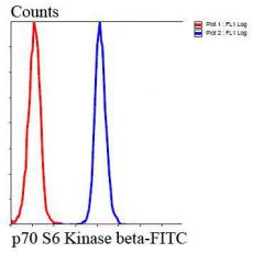
Fig9: Flow cytometric analysis of HepG2 cells with p70 S6 Kinase beta antibody at 1/100 dilution (blue) compared with an unlabelled control (cells without incubation with primary antibody; red). Goat anti rabbit IgG (FITC) was used as the secondary antibo
特别提示:本公司的所有产品仅可用于科研实验,严禁用于临床医疗及其他非科研用途!


