


-

专业包装 正品保证
-

快乐服务 售后无忧
-

会员特权 优惠不断
-

个人信息 严格保护
| 货号 | 规格 | 可用库存 | 销售价(RMB) | 您的折扣价(RMB) | 购买数量 |
|---|
| 熔点: | |
|---|---|
| 密度: | |
| 储存条件: | -20℃ |
Anti-Orai3 antibody
产品描述ORAI3 (ORAI Calcium Release-Activated Calcium Modulator 3) is a Protein Coding gene. Among its related pathways are Calcium signaling pathway. Gene Ontology (GO) annotations related to this gene include store-operated calcium channel activity. An important paralog of this gene is ORAI2.
产品名称Anti-Orai3 antibody
分子量31 kDa
种属反应性Human,Mouse,Rat
验证应用WB,ICC,IHC-P,FC
抗体类型兔多抗
免疫原Synthetic peptide within human Orai-3 aa 10-50.
偶联Non-conjugated
形态Liquid
浓度1 mg/mL.
存放说明Store at +4℃ after thawing. Aliquot store at -20℃. Avoid repeated freeze / thaw cycles.
存储缓冲液1*PBS (pH7.4), 0.2% BSA, 50% Glycerol. Preservative: 0.05% Sodium Azide.
亚型IgG
纯化方式Peptide affinity purified.
亚细胞定位Membrane. Multi-pass membrane protein.
其它名称
WB: 1:500-1:1,000
ICC: 1:50-1:200
IHC-P: 1:50-1:200
FC: 1:50-1:100

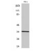
Fig1: Western blot analysis of Orai3 using anti-Orai3 antibody at 1/500 dilution.
Lane 1 : Raji cell lysates
Lane 2 : Raji cell lysates(anti-Orai3 antibody+ immunizing peptide)

Fig2: ICC staining Orai3 in A549 cells (green). The nuclear counter stain is DAPI (blue). Cells were fixed in paraformaldehyde, permeabilised with 0.25% Triton X100/PBS.

Fig3: ICC staining Orai3 in MG-63 cells (green). The nuclear counter stain is DAPI (blue). Cells were fixed in paraformaldehyde, permeabilised with 0.25% Triton X100/PBS.

Fig4: ICC staining Orai3 in SH-SY-5Y cells (green). The nuclear counter stain is DAPI (blue). Cells were fixed in paraformaldehyde, permeabilised with 0.25% Triton X100/PBS.

Fig5: Immunohistochemical analysis of paraffin-embedded rat brain tissue using anti-Orai3 antibody. Counter stained with hematoxylin.

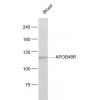
Fig6: Immunohistochemical analysis of paraffin-embedded human liver cancer tissue using anti-Orai3 antibody. Counter stained with hematoxylin.

Fig7: Immunohistochemical analysis of paraffin-embedded mouse testis tissue using anti-Orai3 antibody. Counter stained with hematoxylin.

Fig8: Immunohistochemical analysis of paraffin-embedded human kidney tissue using anti-Orai3 antibody. Counter stained with hematoxylin.

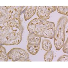
Fig9: Immunohistochemical analysis of paraffin-embedded human placenta tissue using anti-Orai3 antibody. Counter stained with hematoxylin.

Fig10: Flow cytometric analysis of A549 cells with Orai3 antibody at 1/100 dilution (fuchsia) compared with an unlabelled control (cells without incubation with primary antibody; yellow). Alexa Fluor 488-conjugated goat anti-rabbit IgG was used as the sec
特别提示:本公司的所有产品仅可用于科研实验,严禁用于临床医疗及其他非科研用途!


