Anti-macroH2A.1 antibody
-
概述
- 产品描述Variant histone H2A which replaces conventional H2A in a subset of nucleosomes where it represses transcription. Nucleosomes wrap and compact DNA into chromatin, limiting DNA accessibility to the cellular machineries which require DNA as a template. Histones thereby play a central role in transcription regulation, DNA repair, DNA replication and chromosomal stability. DNA accessibility is regulated via a complex set of post-translational modifications of histones, also called histone code, and nucleosome remodeling. Involved in stable X chromosome inactivation. Inhibits the binding of transcription factors, including NF-kappa-B, and interferes with the activity of remodeling SWI/SNF complexes. Inhibits histone acetylation by EP300 and recruits class I HDACs, which induces a hypoacetylated state of chromatin.
- 产品名称Anti-macroH2A.1 antibody
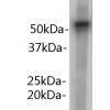
- 分子量40 kDa
- 种属反应性Human,Mouse
- 验证应用WB,IHC-P,FC
- 抗体类型兔多抗
- 免疫原Recombinant protein within human macroH2A.1 aa 200-400.
- 偶联Non-conjugated
-
性能
- 形态Liquid
- 浓度1 mg/mL.
- 存放说明Store at +4℃ after thawing. Aliquot store at -20℃. Avoid repeated freeze / thaw cycles.
- 存储缓冲液1*PBS (pH7.4), 0.2% BSA, 50% Glycerol. Preservative: 0.05% Sodium Azide.
- 亚型IgG
- 纯化方式Protein affinity purified.
- 亚细胞定位Nucleus.
- 其它名称Core histone macro h2a.1 antibody
Core histone macro-H2A.1 antibody
H2A histone family member Y antibody
H2A.y antibody
H2A/y antibody
H2AF12M antibody
H2AFJ antibody
H2afy antibody
H2AY_HUMAN antibody
Histone H2A.Y antibody
Histone macroH2A1 antibody
Histone macroH2A1.1 antibody
Histone macroH2A1.2 antibody
Macroh2a1 antibody
MACROH2A1.1 antibody
MacroH2A1.2 antibody
Medulloblastoma antigen MU MB 50.205 antibody
Medulloblastoma antigen MU-MB-50.205 antibody
mH2a antibody
mH2A1 antibody
more
-
应用
WB: 1:500
IHC-P: 1:50-1:200
FC: 1:50-1:100
-
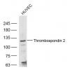
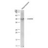
Fig1: Western blot analysis of macroH2A.1 on different lysates using anti-macroH2A.1 antibody at 1/500 dilution.
Positive control:
Lane 1: mouse pancreas
Lane 2: SiHa
Lane 3: PC-3M
Fig2: Immunohistochemical analysis of paraffin-embedded human colon tissue using anti-macroH2A.1 antibody. Counter stained with hematoxylin.
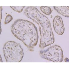
Fig3: Immunohistochemical analysis of paraffin-embedded human placenta tissue using anti-macroH2A.1 antibody. Counter stained with hematoxylin.
Fig4: Immunohistochemical analysis of paraffin-embedded mouse brain tissue using anti-macroH2A.1 antibody. Counter stained with hematoxylin.
Fig5: Flow cytometric analysis of SiHa cells with macroH2A.1 antibody at 1/100 dilution (purple) compared with an unlabelled control (cells without incubation with primary antibody; yellow). Alexa Fluor 488-conjugated goat anti-rabbit IgG was used as the secondary antibody.
特别提示:本公司的所有产品仅可用于科研实验,严禁用于临床医疗及其他非科研用途!