Anti-SHP-1 antibody
-
概述
- 产品描述The steady state of protein tyrosyl phosphorylation in cells is regulated by the opposing action of tyrosine kinases and protein tyrosine phosphatases (PTPs). Several groups have independently identified a non-transmembrane PTP, designated SH-PTP1 (also known as PTP1C, HCP and SHP), which is primarily expressed in hematopoietic cells and characterized by the presence of two SH2 domains N-terminal to the PTP domain. SH2 domains generally mediate the association of regulatory molecules with specific phosphotyrosine-containing sites on autophosphorylated receptors, thereby controlling the initial interaction of receptors with these substrates. A second and much more widely expressed PTP with SH2 domains, SH-PTP2 (also designated PTP1D and Syp), has been identified. Strong sequence similarity between SH-PTP2 and the Drosophila gene corkscrew (CSW) and their similar patterns of expression suggest that SH-PTP2 is the human corkscrew homolog.
- 产品名称Anti-SHP-1 antibody
- 分子量68 kDa
- 种属反应性Human,Mouse,Rat
- 验证应用WB,ICC,IHC-P
- 抗体类型兔多抗
- 免疫原Synthetic peptide of C-terminal human SHP-1.
- 偶联Non-conjugated
-
性能
- 形态Liquid
- 浓度1 mg/mL.
- 存放说明Store at +4℃ after thawing. Aliquot store at -20℃. Avoid repeated freeze / thaw cycles.
- 存储缓冲液1*PBS (pH7.4), 0.2% BSA, 50% Glycerol. Preservative: 0.05% Sodium Azide.
- 亚型IgG
- 纯化方式Peptide affinity purified.
- 亚细胞定位Nucleus. Cytoplasm.
- 其它名称70Z-SHP antibody
EC 3.1.3.48 antibody
HCP antibody
HCPH antibody
Hematopoietic cell phosphatase antibody
Hematopoietic cell protein tyrosine phosphatase antibody
Hematopoietic cell protein-tyrosine phosphatase antibody
HPTP1C antibody
Protein tyrosine phosphatase 1C antibody
Protein tyrosine phosphatase non receptor type 6 antibody
Protein tyrosine phosphatase SHP1 antibody
Protein-tyrosine phosphatase 1C antibody
protein-tyrosine phosphatase SHP 1 antibody
Protein-tyrosine phosphatase SHP-1 antibody
PTN6_HUMAN antibody
PTP 1C antibody
PTP-1C antibody
PTP1C antibody
Ptpn6 antibody
SH PTP 1 antibody
SH PTP1 antibody
SH-PTP1 antibody
SHP 1 antibody
SHP 1L antibody
SHP1 antibody
SHP1L antibody
tyrosine protein phosphatase non receptor type 6 antibody
Tyrosine-protein phosphatase non-receptor type 6 antibody
more
-
应用
WB: 1:500-1:1,000
ICC: 1:50-1:200
IHC-P: 1:50-1:200
-
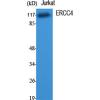
Fig1: Western blot analysis of SHP1 on different lysates using anti-SHP1 antibody at 1/1,000 dilution.
Positive control:
Lane 1: Mouse spleen tissue
Lane 2: HL-60
Lane 3: Rat spleen tissue
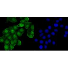
Fig2: ICC staining SHP1 in HepG2 cells (green). The nuclear counter stain is DAPI (blue). Cells were fixed in paraformaldehyde, permeabilised with 0.25% Triton X100/PBS.
Fig3: ICC staining SHP1 in LOVO cells (green). The nuclear counter stain is DAPI (blue). Cells were fixed in paraformaldehyde, permeabilised with 0.25% Triton X100/PBS.
Fig4: Immunohistochemical analysis of paraffin-embedded human spleen tissue using anti- SHP1 antibody. Counter stained with hematoxylin.
Fig5: Immunohistochemical analysis of paraffin-embedded rat spleen tissue using anti- SHP1 antibody. Counter stained with hematoxylin.
Fig6: Immunohistochemical analysis of paraffin-embedded human tonsil tissue using anti- SHP1 antibody. Counter stained with hematoxylin.
特别提示:本公司的所有产品仅可用于科研实验,严禁用于临床医疗及其他非科研用途!