Anti-Raptor antibody
-
概述
- 产品描述Regulatory associated protein of FRAP, also designated Raptor, is a binding partner for mammalian target of rapamycin kinase (FRAP), and is essential for FRAP signalling in vivo. Raptor binding to FRAP is critical for FRAP-catalysed substrate phosphorylation of 4E-BP1. The raptor-FRAP complex is nutrient-sensitive and is important for a mechanism by which cells coordinate cell growth and size with changing environmental conditions. Raptor serves as a negative regulator of FRAP kinase activity under nutrient-deprived conditions and is an important component in the FRAP pathway. Raptor is highly expressed in skeletal muscle and to a lesser extent in brain, kidney, lung and placenta.
- 产品名称Anti-Raptor antibody
- 分子量131 kDa
- 种属反应性Human,Mouse,Rat
- 验证应用WB,ICC,IHC-P,FC
- 抗体类型兔多抗
- 免疫原Synthetic peptide within human Raptor aa 100-150.
- 偶联Non-conjugated
-
性能
- 形态Liquid
- 浓度1 mg/mL.
- 存放说明Store at +4℃ after thawing. Aliquot store at -20℃. Avoid repeated freeze / thaw cycles.
- 存储缓冲液1*PBS (pH7.4), 0.2% BSA, 50% Glycerol. Preservative: 0.05% Sodium Azide.
- 亚型IgG
- 纯化方式Peptide affinity purified.
- 亚细胞定位Cytoplasm. Lysosome.
- 其它名称KIAA1303 antibody
KOG1 antibody
Mip1 antibody
P150 target of rapamycin (TOR) scaffold protein antibody
p150 target of rapamycin (TOR) scaffold protein containing WD repeats antibody
P150 target of rapamycin (TOR)-scaffold protein antibody
Raptor antibody
Regulatory associated protein of mTOR antibody
Regulatory associated protein of MTOR complex 1 antibody
Regulatory-associated protein of mTOR antibody
RPTOR antibody
RPTOR_HUMAN antibody
more
-
应用
WB: 1:500
ICC: 1:50-1:200
IHC-P: 1:50-1:200
FC: 1:50-1:100
-
Fig1: Western blot analysis of Raptor on MCF-7 cell lysate using anti-Raptor antibody at 1/500 dilution.
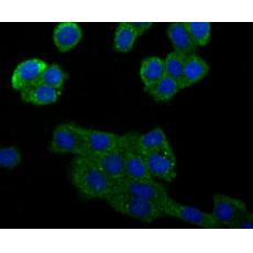
Fig2: ICC staining Raptor in 293T cells (green). The nuclear counter stain is DAPI (blue). Cells were fixed in paraformaldehyde, permeabilised with 0.25% Triton X100/PBS.
Fig3: ICC staining Raptor in LOVO cells (green). The nuclear counter stain is DAPI (blue). Cells were fixed in paraformaldehyde, permeabilised with 0.25% Triton X100/PBS.
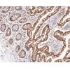
Fig4: Immunohistochemical analysis of paraffin-embedded rat brain tissue using anti-Raptor antibody. Counter stained with hematoxylin.
Fig5: Immunohistochemical analysis of paraffin-embedded human breast tissue using anti-Raptor antibody. Counter stained with hematoxylin.
Fig6: Immunohistochemical analysis of paraffin-embedded mouse colon tissue using anti-Raptor antibody. Counter stained with hematoxylin.
Fig7: Flow cytometric analysis of A549 cells with Raptor antibody at 1/100 dilution (red) compared with an unlabelled control (cells without incubation with primary antibody; black). Alexa Fluor 488-conjugated goat anti-rabbit IgG was used as the secondar
特别提示:本公司的所有产品仅可用于科研实验,严禁用于临床医疗及其他非科研用途!