Anti-DIAPH3 antibody
-
概述
- 产品描述DIAPH3 (diaphanous homolog 3), also known as DIAP3, DRF3 or mDia2 of mouse origin, is a 1,193 amino acid member of the formin homology protein family and is required for the correct function of various cellular processes. DIAPH3 binds to both Profilin, a protein involved in cell maintenance, and to the GTP-bound form of Rho (Rho-GTP). Binding to both of these proteins allows DIAPH3 to recruit Profilin to the membrane, in a Rho-dependent manner. At the membrane, DIAPH3 promotes Actin polymerization and is required for stress fiber formation, cytokinesis and transcriptional activation of the serum response factor (SRF). DIAPH3 also regulates Actin dynamics by coupling Src tyrosine kinase (c-Src) and Rho during Actin signaling events. DIAPH3 contains one diaph-anous autoregulatory domain (DAD) and one Rho GTPase-binding domain (GBD). When DAD and GBD are intramolecularly bound, the GBD is occupied and DIAPH3 is inactive. Interruption of the DAD-GBD bond allows the GBD to bind to Rho-GTP, thus activating DIAPH3. Seven isoforms of DIAPH3 exist due to alternative splicing events.
- 产品名称Anti-DIAPH3 antibody
- 分子量137/80 kDa
- 种属反应性Human,Mouse
- 验证应用WB,ICC,IHC-P,FC
- 抗体类型兔多抗
- 免疫原Synthetic peptide within human DIAPH3 aa 1-50.
- 偶联Non-conjugated
-
性能
- 形态Liquid
- 浓度1 mg/mL.
- 存放说明Store at +4℃ after thawing. Aliquot store at -20℃. Avoid repeated freeze / thaw cycles.
- 存储缓冲液1*PBS (pH7.4), 0.2% BSA, 50% Glycerol. Preservative: 0.05% Sodium Azide.
- 亚型IgG
- 纯化方式Peptide affinity purified.
- 亚细胞定位Cytosol.
- 其它名称AN antibody
AUNA1 antibody
Dia2 antibody
diap3 antibody
DIAP3_HUMAN antibody
DIAPH3 antibody
Diaphanous homolog 3 (Drosophila) antibody
Diaphanous homolog 3 antibody
Diaphanous related formin 3 antibody
Diaphanous, Drosophila, homolog of, 3 antibody
Diaphanous-related formin-3 antibody
DKFZp434C0931 antibody
DKFZp686A13178 antibody
DRF3 antibody
FLJ34705 antibody
mDia2 antibody
NSDAN antibody
OTTHUMP00000018480 antibody
Protein diaphanous homolog 3 antibody
RP11-26P21.1 antibody
more
-
应用
ICC: 1:50-1:200
IHC-P: 1:50-1:200
FC: 1:50-1:100
WB: 1:500
-
Fig1: Western blot analysis of DIAPH3 on SiHa cell lysate using anti-DIAPH3 antibody at 1/500 dilution.
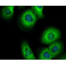
Fig2: ICC staining DIAPH3 in LOVO cells (green). The nuclear counter stain is DAPI (blue). Cells were fixed in paraformaldehyde, permeabilised with 0.25% Triton X100/PBS.
Fig3: ICC staining DIAPH3 in SiHa cells (green). The nuclear counter stain is DAPI (blue). Cells were fixed in paraformaldehyde, permeabilised with 0.25% Triton X100/PBS.
Fig4: ICC staining DIAPH3 in A431 cells (green). The nuclear counter stain is DAPI (blue). Cells were fixed in paraformaldehyde, permeabilised with 0.25% Triton X100/PBS.
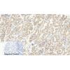
Fig5: Immunohistochemical analysis of paraffin-embedded human placenta tissue using anti-DIAPH3 antibody. Counter stained with hematoxylin.
Fig6: Immunohistochemical analysis of paraffin-embedded human kidney tissue using anti-DIAPH3 antibody. Counter stained with hematoxylin.
Fig7: Flow cytometric analysis of LOVO cells with DIAPH3 antibody at 1/100 dilution (red) compared with an unlabelled control (cells without incubation with primary antibody; black). Alexa Fluor 488-conjugated goat anti-rabbit IgG was used as the secondar
特别提示:本公司的所有产品仅可用于科研实验,严禁用于临床医疗及其他非科研用途!


