Anti-Hip1 antibody
-
概述
- 产品描述Huntington disease is associated with the expansion of a polyglutamine tract, greater than 35 repeats, in the HD gene product huntingtin. HIP1 (huntingtin-interacting protein 1), a membrane-associated protein, binds specifically to the N-terminus of human huntingtin. HIP1 is ubiquitously expressed in different brain regions at low levels, and exhibits nearly identical subcellular fractionation as huntingtin. The huntingtin-HIP1 interaction is restricted to the brain and is inversely correlated to the polyglutamine length in the huntingtin, suggesting that loss of normal huntingtin-HIP1 interaction may compromise the membrane-cytoskeletal integrity in the brain. HIP1 contains an endocytic multidomain protein with a C-terminal Actin-binding domain, a central coiled-coil forming region and an N-terminal ENTH domain. HIP1 may be involved in vesicle trafficking; the structural integrity of HIP1 is crucial for maintenance of normal vesicle size in vivo. HIP12 is a non-proapoptotic member of the HIP gene family that is expressed in the brain and shares a similar subcellular distribution pattern with HIP1. However, HIP12 differs from HIP1 in its pattern of expression at both the mRNA and protein level. HIP12 does not directly interact with huntingtin but can interact with HIP1.
- 产品名称Anti-Hip1 antibody
- 分子量116 kDa
- 种属反应性Human,Mouse,Rat
- 验证应用WB,ICC,IHC-P,FC
- 抗体类型兔多抗
- 免疫原Synthetic peptide within C terminal human Hip1.
- 偶联Non-conjugated
-
性能
- 形态Liquid
- 浓度1 mg/mL.
- 存放说明Store at +4℃ after thawing. Aliquot store at -20℃. Avoid repeated freeze / thaw cycles.
- 存储缓冲液1*PBS (pH7.4), 0.2% BSA, 50% Glycerol. Preservative: 0.05% Sodium Azide.
- 亚型IgG
- 纯化方式Peptide affinity purified.
- 亚细胞定位Nucleus. Cytoplasm.
- 其它名称2610109B09Rik antibody
A930014B11Rik antibody
E130315I21Rik antibody
HIP 1 antibody
HIP I antibody
HIP-1 antibody
HIP-I antibody
hip1 antibody
HIP1/PDGFRB fusion gene antibody
HIP1/PDGFRB fusion gene, included antibody
HIP1_HUMAN antibody
HIPI antibody
Huntingtin interacting protein 1 antibody
Huntingtin-interacting protein 1 antibody
Huntingtin-interacting protein I antibody
ILWEQ antibody
KIAA4113 antibody
MGC126506 antibody
MGC27616 antibody
mKIAA4113 antibody
more
-
应用
WB: 1:500-1:1,000
ICC: 1:100-1:500
IHC-P: 1:50-1:200
FC: 1:50-1:100
-
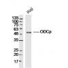
Fig1: Western blot analysis of Hip1 on different tissue lysates using anti-Hip1 antibody at 1/500 dilution.
Positive control:
Lane 1: Mouse testis
Lane 2: Mouse spinal cord
Lane 3: SH-SY5Y
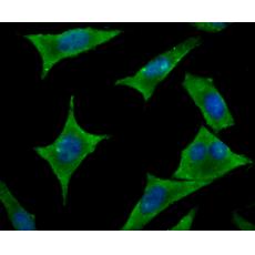
Fig2: ICC staining Hip1 in A549 cells (green). The nuclear counter stain is DAPI (blue). Cells were fixed in paraformaldehyde, permeabilised with 0.25% Triton X100/PBS.
Fig3: ICC staining Hip1 in PC-3M cells (green). The nuclear counter stain is DAPI (blue). Cells were fixed in paraformaldehyde, permeabilised with 0.25% Triton X100/PBS.
Fig4: ICC staining Hip1 in SH-SY-5Y cells (green). The nuclear counter stain is DAPI (blue). Cells were fixed in paraformaldehyde, permeabilised with 0.25% Triton X100/PBS.
Fig5: Immunohistochemical analysis of paraffin-embedded human colon cancer tissue using anti-Hip1 antibody. Counter stained with hematoxylin.
Fig6: Immunohistochemical analysis of paraffin-embedded mouse brain tissue using anti-Hip1 antibody. Counter stained with hematoxylin.
Fig7: Immunohistochemical analysis of paraffin-embedded mouse fallopian tubes tissue using anti-Hip1 antibody. Counter stained with hematoxylin.
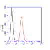
Fig8: Flow cytometric analysis of SH-SY5Y cells with Hip1 antibody at 1/100 dilution (red) compared with an unlabelled control (cells without incubation with primary antibody; black). Alexa Fluor 488-conjugated goat anti-rabbit IgG was used as the seconda
特别提示:本公司的所有产品仅可用于科研实验,严禁用于临床医疗及其他非科研用途!