Anti-Vitamin D Receptor antibody
-
概述
- 产品描述The active metabolite of vitamin D modulates the expression of a wide variety of genes in a developmentally specific manner. This secosteroid hormone can up- or downregulate the expression of genes involved in a diverse array of responses such as proliferation, differentiation and calcium homeostasis. 1,25-(OH)2-vitamin D3 exerts its effects through interaction with the vitamin D receptor (VDR), a member of the superfamily of hormone-activated nuclear receptors. In its ligand-bound state, the VDR forms heterodimers with the 9-cis retinoic acid receptor, RXR, and affects gene expression by binding specific DNA sequences known as hormone response elements, or HREs. In addition to regulating the above-mentioned cellular responses, 1,25-(OH)2-vitamin D3 exhibits antiproliferative properties in osteosarcoma, melanoma, colon carcinoma and breast carcinoma cells.
- 产品名称Anti-Vitamin D Receptor antibody
- 分子量48 kDa
- 种属反应性Human
- 验证应用WB,FC
- 抗体类型兔多抗
- 免疫原Peptide
- 偶联Non-conjugated
-
性能
- 形态Liquid
- 浓度1 mg/mL.
- 存放说明Store at +4℃ after thawing. Aliquot store at -20℃ or -80℃. Avoid repeated freeze / thaw cycles.
- 存储缓冲液1*PBS (pH7.4), 0.2% BSA, 50% Glycerol. Preservative: 0.05% Sodium Azide.
- 亚型IgG
- 纯化方式Peptide affinity purified
- 亚细胞定位Nucleus. Cytoplasm.
- 其它名称1 25 dihydroxyvitamin D3 receptor antibody
1 antibody
1,25 dihydroxyvitamin D3 receptor antibody
1,25-@dihydroxyvitamin D3 receptor antibody
25-dihydroxyvitamin D3 receptor antibody
Member 1 antibody
NR1I1 antibody
Nuclear receptor subfamily 1 group I member 1 antibody
PPP1R163 antibody
Protein phosphatase 1, regulatory subunit 163 antibody
VDR antibody
VDR_HUMAN antibody
Vitamin D (1,25- dihydroxyvitamin D3) receptor antibody
Vitamin D hormone receptor antibody
Vitamin D nuclear receptor variant 1 antibody
Vitamin D receptor antibody
Vitamin D3 receptor antibody
more
-
应用
WB: 1:500-1:1,000
FC: 1:50-1:100
-
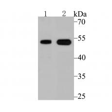
Fig1: Western blot analysis of Vitamin D Receptor on PC-3M cell and human small intestine tissue lysate using anti-Vitamin D Receptor antibody at 1/500 dilution.
Fig2: Flow cytometric analysis of LOVO cells with Vitamin D Receptor antibody at 1/100 dilution (red) compared with an unlabelled control (cells without incubation with primary antibody; black). Alexa Fluor 488-conjugated goat anti-rabbit IgG was used as the secondary antibody.
特别提示:本公司的所有产品仅可用于科研实验,严禁用于临床医疗及其他非科研用途!


