Anti-SATB1 antibody
-
概述
- 产品描述The homeoproteins CCAAT displacement protein (CDP) and special AT-rich sequence binding protein 1 (SATB1) are transcriptional repressors of many cellular genes, and they participate in cell development and cell type differentiation. SATB1 is expressed primarily in thymocytes, and, like CDP, it also contains a distinct homeobox DNA-binding domain that is essential for DNA binding. SATB1 and CDP interact through these homeodomains and synergistically function as mediators of gene expression. SATB1 contains an additional domain that has a higher affinity for DNA and specifically facilitates the direct association between SATB1 and the nuclear matrix attachment regions (MARs) of DNA. MARs are specific DNA sequences that bind to the nuclear matrix and form the base of chromosomal loops that organize the chromosomes and regulate DNA transcription and replication within the nucleus. The association of SATB1 with the core unwinding element within the base-unpairing region of MARs requires both the MAR and homeobox binding domains of SATB1.
- 产品名称Anti-SATB1 antibody
- 分子量Predicted band size 86 kDa
- 种属反应性Human,Mouse
- 验证应用WB,ICC,FC
- 抗体类型兔多抗
- 免疫原Peptide
- 偶联Non-conjugated
-
性能
- 形态Liquid
- 浓度1 mg/mL.
- 存放说明Store at +4℃ after thawing. Aliquot store at -20℃ or -80℃. Avoid repeated freeze / thaw cycles.
- 存储缓冲液1*PBS (pH7.4), 0.2% BSA, 50% Glycerol. Preservative: 0.05% Sodium Azide.
- 亚型IgG
- 纯化方式Peptide affinity purified
- 亚细胞定位Nucleus.
- 其它名称DNA binding protein SATB1 antibody
DNA-binding protein SATB1 antibody
SATB homeobox 1 antibody
SATB1 antibody
SATB1_HUMAN antibody
Special AT rich sequence binding protein 1 (binds to nuclear matrix/scaffold associating DNA) antibody
Special AT rich sequence binding protein 1 antibody
Special AT-rich sequence-binding protein 1 antibody
more
-
应用
WB: 1:500
ICC: 1:100
FC: 1:50-1:100
-
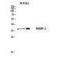
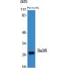
Fig1: Western blot analysis of SATB1 on mouse thymus tissue lysate using anti-SATB1 antibody at 1/500 dilution.
Fig2: ICC staining SATB1 in SH-SY-5Y cells (green). The nuclear counter stain is DAPI (blue). Cells were fixed in paraformaldehyde, permeabilised with 0.25% Triton X100/PBS.
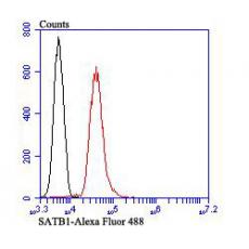
Fig3: Flow cytometric analysis of Jurkat cells with SATB1 antibody at 1/100 dilution (red) compared with an unlabelled control (cells without incubation with primary antibody; black). Alexa Fluor 488-conjugated goat anti rabbit IgG was used as the secondary antibody.
特别提示:本公司的所有产品仅可用于科研实验,严禁用于临床医疗及其他非科研用途!


