Anti-TNFAIP3 antibody
-
概述
- 产品描述A20 is a Cys2/Cys2 zinc finger protein that is induced by a variety of inflammatory stimuli and regulates gene expression. Specifically, A20 is induced by tumor necrosis factor (TNF) and interleukin 1 (IL-1), and acts as a negative regulator of nuclear factor κ B (NFκB) gene expression. By inhibiting NFκB activation, A20 plays a critical role in terminating NFκB responses to various stimuli. Although the C-terminal region of A20 contains seven zinc finger domains, only four of these domains are required for in vitro inhibition of TNF-induced NFκB activation. A20 also interacts with several other proteins, such as TRAF2, TRAF6 and IkB kinase (IKK) γ protein, and can thereby inhibit cell death. TXBP151, a novel A20-binding protein, may mediate the anti-apoptotic activity of A20. Involved in the negative feedback regulation of signal transduction, A20 and A20-binding proteins may be useful as novel therapeutic tools in the treatment of a variety of diseases.
- 产品名称Anti-TNFAIP3 antibody
- 分子量90 kDa
- 种属反应性Human,Mouse,Rat
- 验证应用WB,ICC,IHC-P,FC
- 抗体类型兔多抗
- 免疫原Recombinant protein
- 偶联Non-conjugated
-
性能
- 形态Liquid
- 浓度1 mg/mL.
- 存放说明Store at +4℃ after thawing. Aliquot store at -20℃ or -80℃. Avoid repeated freeze / thaw cycles.
- 存储缓冲液1*PBS (pH7.4), 0.2% BSA, 50% Glycerol. Preservative: 0.05% Sodium Azide.
- 亚型IgG
- 纯化方式Protein A purified.
- 亚细胞定位Cytoplasm. Nucleus.
- 其它名称A20 antibody
AISBL antibody
MGC104522 antibody
MGC138687 antibody
MGC138688 antibody
OTU domain containing protein 7C antibody
OTU domain-containing protein 7C antibody
OTUD7C antibody
Putative DNA binding protein A20 antibody
Putative DNA-binding protein A20 antibody
TNAP3_HUMAN antibody
TNF alpha-induced protein 3 antibody
TNFA1P2 antibody
TNFAIP 3 antibody
TNFAIP3 (A20) antibody
TNFAIP3 antibody
Tumor necrosis factor alpha induced protein 3 antibody
Tumor necrosis factor alpha-induced protein 3 antibody
Tumor necrosis factor induced protein 3 antibody
Tumor necrosis factor inducible protein A20 antibody
tumor necrosis factor, alpha-induced protein 3 antibody
Zinc finger protein A20 antibody
more
-
应用
WB: 1:500
ICC: 1:100-1:500
IHC-P: 1:50-1:200
FC: 1:50-1:100
-
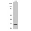
Fig1: Western blot analysis of TNFAIP3 on Jurkat (1) and Daudi (2) cell lysate using anti-TNFAIP3 antibody at 1/100 dilution.
Fig2: ICC staining TNFAIP3 in A431 cells (green). The nuclear counter stain is DAPI (blue). Cells were fixed in paraformaldehyde, permeabilised with 0.25% Triton X100/PBS.
Fig3: ICC staining TNFAIP3 in LOVO cells (green). The nuclear counter stain is DAPI (blue). Cells were fixed in paraformaldehyde, permeabilised with 0.25% Triton X100/PBS.
Fig4: Immunohistochemical analysis of paraffin-embedded rat lung tissue using anti-TNFAIP3 antibody. Counter stained with hematoxylin.
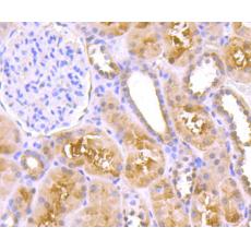
Fig5: Immunohistochemical analysis of paraffin-embedded human kidney tissue using anti-TNFAIP3 antibody. Counter stained with hematoxylin.
Fig6: Immunohistochemical analysis of paraffin-embedded human kidney tissue using anti-TNFAIP3 antibody. Counter stained with hematoxylin.
Fig7: Immunohistochemical analysis of paraffin-embedded mouse testis tissue using anti-TNFAIP3 antibody. Counter stained with hematoxylin.
Fig8: Flow cytometric analysis of HepG2 cells with TNFAIP3 antibody at 1/100 dilution (red) compared with an unlabelled control (cells without incubation with primary antibody; black). Goat anti rabbit IgG (FITC) was used as the secondary antibody.
特别提示:本公司的所有产品仅可用于科研实验,严禁用于临床医疗及其他非科研用途!