Anti-EGFR antibody
-
概述
- 产品描述Receptor tyrosine kinase binding ligands of the EGF family and activating several signaling cascades to convert extracellular cues into appropriate cellular responses. Known ligands include EGF, TGFA/TGF-alpha, amphiregulin, epigen/EPGN, BTC/betacellulin, epiregulin/EREG and HBEGF/heparin-binding EGF. Ligand binding triggers receptor homo- and/or heterodimerization and autophosphorylation on key cytoplasmic residues. The phosphorylated receptor recruits adapter proteins like GRB2 which in turn activates complex downstream signaling cascades. Activates at least 4 major downstream signaling cascades including the RAS-RAF-MEK-ERK, PI3 kinase-AKT, PLCgamma-PKC and STATs modules. May also activate the NF-kappa-B signaling cascad.
- 产品名称Anti-EGFR antibody
- 分子量175 kDa
- 种属反应性Human,Mouse
- 验证应用WB,ICC,IHC-P,FC
- 抗体类型兔多抗
- 免疫原Peptide.
- 偶联Non-conjugated
-
性能
- 形态Liquid
- 浓度1 mg/mL.
- 存放说明Store at +4℃ after thawing. Aliquot store at -20℃ or -80℃. Avoid repeated freeze / thaw cycles.
- 存储缓冲液1*PBS (pH7.4), 0.2% BSA, 50% Glycerol. Preservative: 0.05% Sodium Azide.
- 亚型IgG
- 纯化方式Peptide affinity purified
- 亚细胞定位Plasma membrane, Nucleus, Endosome, Endoplasmic reticulum.
- 其它名称Avian erythroblastic leukemia viral (v erb b) oncogene homolog antibody
Cell growth inhibiting protein 40 antibody
Cell proliferation inducing protein 61 antibody
EGF R antibody
EGFR antibody
EGFR_HUMAN antibody
Epidermal growth factor receptor (avian erythroblastic leukemia viral (v erb b) oncogene homolog) antibody
Epidermal growth factor receptor (erythroblastic leukemia viral (v erb b) oncogene homolog avian) antibody
Epidermal growth factor receptor antibody
erb-b2 receptor tyrosine kinase 1 antibody
ERBB antibody
ERBB1 antibody
Errp antibody
HER1 antibody
mENA antibody
NISBD2 antibody
Oncogen ERBB antibody
PIG61 antibody
Proto-oncogene c-ErbB-1 antibody
Receptor tyrosine protein kinase ErbB 1 antibody
Receptor tyrosine-protein kinase ErbB-1 antibody
SA7 antibody
Species antigen 7 antibody
Urogastrone antibody
v-erb-b Avian erythroblastic leukemia viral oncogen homolog antibody
wa2 antibody
Wa5 antibody
more
-
应用
WB: 1:500-2,000
ICC: 1:50-1:200
IHC-P: 1:50-1:200
FC: 1:50-1:100
-
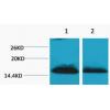
Fig1: Western blot analysis of EGFR on HepG2 (1) and Hela (2) cell lysates using anti-EGFR antibody at 1/1,000 dilution.
Fig2: ICC staining EGFR in LOVO cells (green). The nuclear counter stain is DAPI (blue). Cells were fixed in paraformaldehyde, permeabilised with 0.25% Triton X100/PBS.
Fig3: ICC staining EGFR in SW480 cells (green). The nuclear counter stain is DAPI (blue). Cells were fixed in paraformaldehyde, permeabilised with 0.25% Triton X100/PBS.
Fig4: Immunohistochemical analysis of paraffin-embedded human liver tissue using anti-EGFR antibody. Counter stained with hematoxylin.
Fig5: Immunohistochemical analysis of paraffin-embedded human kidney tissue using anti-EGFR antibody. Counter stained with hematoxylin.
Fig6: Immunohistochemical analysis of paraffin-embedded human placenta tissue using anti-EGFR antibody. Counter stained with hematoxylin.
Fig7: Flow cytometric analysis of A431 cells with EGFR antibody at 1/100 dilution (red) compared with an unlabelled control (cells without incubation with primary antibody; black). Alexa Fluor 488-conjugated goat anti-rabbit IgG was used as the secondary
特别提示:本公司的所有产品仅可用于科研实验,严禁用于临床医疗及其他非科研用途!