Anti-CDH17 antibody
-
概述
- 产品描述This gene is a member of the cadherin superfamily, genes encoding calcium-dependent, membrane-associated glycoproteins. The encoded protein is cadherin-like, consisting of an extracellular region, containing 7 cadherin domains, and a transmembrane region but lacking the conserved cytoplasmic domain. The protein is a component of the gastrointestinal tract and pancreatic ducts, acting as an intestinal proton-dependent peptide transporter in the first step in oral absorption of many medically important peptide-based drugs. The protein may also play a role in the morphological organization of liver and intestine. Alternative splicing results in multiple transcript variants.
- 产品名称Anti-CDH17 antibody
- 分子量Predicted band size: 92 kDa
- 种属反应性Human,Mouse,Rat
- 验证应用WB,IHC-P,ICC
- 抗体类型兔多抗
- 免疫原Recombinant protein within Human CDH17 aa 33-210.
- 偶联Non-conjugated
-
性能
- 形态Liquid
- 浓度1 mg/mL.
- 存放说明Store at +4℃ after thawing. Aliquot store at -20℃. Avoid repeated freeze / thaw cycles.
- 存储缓冲液1*PBS (pH7.4), 0.2% BSA, 50% Glycerol. Preservative: 0.05% Sodium Azide.
- 亚型IgG
- 纯化方式Protein affinity purified.
- 亚细胞定位Cell membrane.
- 其它名称BILL-cadherin antibody
CAD17_HUMAN antibody
Cadherin 16 antibody
Cadherin 16, formerly antibody
Cadherin 17 antibody
cadherin 17, LI cadherin (liver-intestine) antibody
cadherin, liver-intestine antibody
Cadherin-17 antibody
CDH16 antibody
CDH16, formerly antibody
Cdh17 antibody
FLJ26931 antibody
Formerly Cadherin 16 antibody
Formerly CDH16 antibody
HPT 1 antibody
HPT-1 cadherin antibody
human intestinal peptide-associated transporter HPT-1 antibody
human peptide transporter 1 antibody
Intestinal peptide-associated transporter HPT 1 antibody
Intestinal peptide-associated transporter HPT-1 antibody
LI-cadherin antibody
Liver Cadherin antibody
liver intestine cadherin antibody
Liver-intestine cadherin antibody
more
-
应用
WB:1:500-1:1,000
ICC:1:50-1:200
IHC-P:1:50-1:200
-
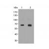
Fig1: Western blot analysis of CDH17 on human colon tissue lysate. Proteins were transferred to a PVDF membrane and blocked with 5% BSA in PBS for 1 hour at room temperature. The primary antibody was used at a 1:1,000 dilution in 5% BSA at room temperature for 2 hours. Goat Anti-Rabbit IgG - HRP Secondary Antibody (HA1001) at 1:5,000 dilution was used for 1 hour at room temperature.
Fig2: ICC staining of CDH17 in PANC-1 cells (green). Formalin fixed cells were permeabilized with 0.1% Triton X-100 in TBS for 10 minutes at room temperature and blocked with 1% Blocker BSA for 15 minutes at room temperature. Cells were probed with the antibody at a dilution of 1:50 for 1 hour at room temperature, washed with PBS. Alexa Fluor®488 Goat anti-Rabbit IgG was used as the secondary antibody at 1/100 dilution. The nuclear counter stain is DAPI (blue).
Fig3: Immunohistochemical analysis of paraffin-embedded rat colon tissue using anti-CDH17 antibody. The section was pre-treated using heat mediated antigen retrieval with Tris-EDTA buffer (pH 8.0-8.4) for 20 minutes.The tissues were blocked in 5% BSA for 30 minutes at room temperature, washed with ddH2O and PBS, and then probed with the antibodyat 1/50 dilution, for 30 minutes at room temperature and detected using an HRP conjugated compact polymer system. DAB was used as the chromogen. Counter stained with hematoxylin and mounted with DPX.
Fig4: Immunohistochemical analysis of paraffin-embedded human colon tissue using anti-CDH17 antibody. The section was pre-treated using heat mediated antigen retrieval with Tris-EDTA buffer (pH 8.0-8.4) for 20 minutes.The tissues were blocked in 5% BSA for 30 minutes at room temperature, washed with ddH2O and PBS, and then probed with the antibodyat 1/50 dilution, for 30 minutes at room temperature and detected using an HRP conjugated compact polymer system. DAB was used as the chromogen. Counter stained with hematoxylin and mounted with DPX.
Fig5: Immunohistochemical analysis of paraffin-embedded human appendix tissue using anti-CDH17 antibody. The section was pre-treated using heat mediated antigen retrieval with Tris-EDTA buffer (pH 8.0-8.4) for 20 minutes.The tissues were blocked in 5% BSA for 30 minutes at room temperature, washed with ddH2O and PBS, and then probed with the antibody at 1/200 dilution, for 30 minutes at room temperature and detected using an HRP conjugated compact polymer system. DAB was used as the chromogen. Counter stained with hematoxylin and mounted with DPX.
Fig6: Immunohistochemical analysis of paraffin-embedded human stomach cancer tissue using anti-CDH17 antibody. The section was pre-treated using heat mediated antigen retrieval with Tris-EDTA buffer (pH 8.0-8.4) for 20 minutes.The tissues were blocked in 5% BSA for 30 minutes at room temperature, washed with ddH2O and PBS, and then probed with the antibody) at 1/200 dilution, for 30 minutes at room temperature and detected using an HRP conjugated compact polymer system. DAB was used as the chromogen. Counter stained with hematoxylin and mounted with DPX.
Fig7: Immunohistochemical analysis of paraffin-embedded mouse small intestine tissue using anti-CDH17 antibody. The section was pre-treated using heat mediated antigen retrieval with Tris-EDTA buffer (pH 8.0-8.4) for 20 minutes.The tissues were blocked in
特别提示:本公司的所有产品仅可用于科研实验,严禁用于临床医疗及其他非科研用途!