


-

专业包装 正品保证
-

快乐服务 售后无忧
-

会员特权 优惠不断
-

个人信息 严格保护
| 货号 | 规格 | 可用库存 | 销售价(RMB) | 您的折扣价(RMB) | 购买数量 |
|---|
| 熔点: | |
|---|---|
| 密度: | |
| 储存条件: | -20℃ |
Anti-FSH beta antibody
产品描述Follitropin subunit beta also known as follicle-stimulating hormone beta subunit (FSH-B) is a protein that in humans is encoded by the FSHB gene. The pituitary glycoprotein hormone family includes follicle-stimulating hormone, luteinizing hormone, chorionic gonadotropin, and thyroid-stimulating hormone.Alternative splicing results in two transcript variants encoding the same protein. The pituitary glycoprotein hormone family includes follicle-stimulating hormone, luteinizing hormone, chorionic gonadotropin, and thyroid-stimulating hormone. All of these glycoproteins consist of an identical alpha subunit and a hormone-specific beta subunit. This gene encodes the beta subunit of follicle-stimulating hormone. In conjunction with luteinizing hormone, follicle-stimulating hormone induces egg and sperm production.
产品名称Anti-FSH beta antibody
分子量15 kDa (Predicted band size)
种属反应性Human,Mouse,Rat
验证应用WB,IHC-P,FC
抗体类型兔多抗
免疫原Synthetic peptide within human FSH beta aa 40-80.
偶联Non-conjugated
形态Liquid
浓度1 mg/mL
存放说明Store at +4℃ after thawing. Aliquot store at -20℃. Avoid repeated freeze / thaw cycles.
存储缓冲液1*PBS (pH7.4), 0.2% BSA, 50% Glycerol. Preservative: 0.05% Sodium Azide.
亚型IgG
纯化方式Peptide affinity purified.
亚细胞定位Secreted.
其它名称
WB: 1:500-1:2,000
IHC-P: 1:50-1:100
FC: 1:50-1:100

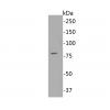
Fig1: Western blot analysis of FSH beta on SH-SY5Y cell lysate. Proteins were transferred to a PVDF membrane and blocked with 5% BSA in PBS for 1 hour at room temperature. The primary antibody was used in 5% BSA at room temperature for 2 hours. Goat Anti-Rabbit IgG - HRP Secondary Antibody (HA1001) at 1:5,000 dilution was used for 1 hour at room temperature.

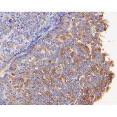
Fig2: Immunohistochemical analysis of paraffin-embedded rat pituitary tissue using anti-FSH beta antibody. The section was pre-treated using heat mediated antigen retrieval with Tris-EDTA buffer (pH 8.0-8.4) for 20 minutes.The tissues were blocked in 5% BSA for 30 minutes at room temperature, washed with ddH2O and PBS, and then probed with the primary antibodyfor 30 minutes at room temperature. The detection was performed using an HRP conjugated compact polymer system. DAB was used as the chromogen. Tissues were counterstained with hematoxylin and mounted with DPX.

Fig3: Immunohistochemical analysis of paraffin-embedded human pituitary tissue using anti-FSH beta antibody. The section was pre-treated using heat mediated antigen retrieval with Tris-EDTA buffer (pH 8.0-8.4) for 20 minutes.The tissues were blocked in 5% BSA for 30 minutes at room temperature, washed with ddH2O and PBS, and then probed with the primary antibody or 30 minutes at room temperature. The detection was performed using an HRP conjugated compact polymer system. DAB was used as the chromogen. Tissues were counterstained with hematoxylin and mounted with DPX.

Fig4: Immunohistochemical analysis of paraffin-embedded human liver tissue using anti-FSH beta antibody. The section was pre-treated using heat mediated antigen retrieval with Tris-EDTA buffer (pH 8.0-8.4) for 20 minutes.The tissues were blocked in 5% BSA for 30 minutes at room temperature, washed with ddH2O and PBS, and then probed with the primary antibodyor 30 minutes at room temperature. The detection was performed using an HRP conjugated compact polymer system. DAB was used as the chromogen. Tissues were counterstained with hematoxylin and mounted with DPX.

Fig5: Immunohistochemical analysis of paraffin-embedded mouse pituitary tissue using anti-FSH beta antibody. The section was pre-treated using heat mediated antigen retrieval with Tris-EDTA buffer (pH 8.0-8.4) for 20 minutes.The tissues were blocked in 5% BSA for 30 minutes at room temperature, washed with ddH2O and PBS, and then probed with the primary antibody for 30 minutes at room temperature. The detection was performed using an HRP conjugated compact polymer system. DAB was used as the chromogen. Tissues were counterstained with hematoxylin and mounted with DPX.

Fig6: Flow cytometric analysis of FSH beta was done on SH-SY5Y cells. The cells were fixed, permeabilized and stained with the primary antibody (red). After incubation of the primary antibody at room temperature for an hour, the cells were stained with a Alexa Fluor 488-conjugated Goat anti-Rabbit IgG Secondary antibody at 1/1000 dilution for 30 minutes.Unlabelled sample was used as a control (cells without incubation with primary antibody; black).
特别提示:本公司的所有产品仅可用于科研实验,严禁用于临床医疗及其他非科研用途!


