


-

专业包装 正品保证
-

快乐服务 售后无忧
-

会员特权 优惠不断
-

个人信息 严格保护
| 别名: | Cytokeratin 17 | ||
|---|---|---|---|
| 适用物种: | Human,Mouse,Rat | ||
| 验证应用: | WB,IHC-P,ICC,FC | ||
| 种属: | 兔多抗 | ||
| 储存条件: | -20℃ | ||
|
| 货号 | 规格 | 可用库存 | 销售价(RMB) | 您的折扣价(RMB) | 购买数量 | ||||||
|---|---|---|---|---|---|---|---|---|---|---|---|
| ZY6Cytokeratin 17R-50 μl | 兔多抗 | 现货 | 1500 | ||||||||
| ZY6Cytokeratin 17R-100 μl | 兔多抗 | 现货 | 2500 | ||||||||
| 熔点: | |
|---|---|
| 密度: | |
| 储存条件: | -20℃ |
Anti-Cytokeratin 17 antibody
产品描述Type I keratin involved in the formation and maintenance of various skin appendages, specifically in determining shape and orientation of hair (By similarity). Required for the correct growth of hair follicles, in particular for the persistence of the anagen (growth) state (By similarity). Modulates the function of TNF-alpha in the specific context of hair cycling. Regulates protein synthesis and epithelial cell growth through binding to the adapter protein SFN and by stimulating Akt/mTOR pathway (By similarity). Involved in tissue repair. May be a marker of basal cell differentiation in complex epithelia and therefore indicative of a certain type of epithelial "stem cells". Acts as a promoter of epithelial proliferation by acting a regulator of immune response in skin: promotes Th1/Th17-dominated immune environment contributing to the development of basaloid skin tumors (By similarity). May act as an autoantigen in the immunopathogenesis of psoriasis, with certain peptide regions being a major target for autoreactive T-cells and hence causing their proliferation.
产品名称Anti-Cytokeratin 17 antibody
分子量48 kDa
种属反应性Human,Mouse,Rat
验证应用WB,IHC-P,ICC,FC
抗体类型兔多抗
免疫原Synthetic peptide within C-terminal Human Cytokeratin 17.
偶联Non-conjugated
形态Liquid
浓度1 mg/mL.
存放说明Store at +4℃ after thawing. Aliquot store at -20℃. Avoid repeated freeze / thaw cycles.
存储缓冲液1*PBS (pH7.4), 0.2% BSA, 50% Glycerol. Preservative: 0.05% Sodium Azide.
亚型IgG
纯化方式Peptide affinity purified.
亚细胞定位Cytoplasm.
其它名称
WB:1:5,000-1:10,000
ICC:1:100-1:400
IHC-P:1:50-1:200
FC:1:50-1:100

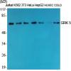
Fig1: Western blot analysis of Cytokeratin 17 on SiHa cell lysate. Proteins were transferred to a PVDF membrane and blocked with 5% BSA in PBS for 1 hour at room temperature. The primary antibody was used in 5% BSA at room temperature for 2 hours. Goat Anti-Rabbit IgG - HRP Secondary Antibody (HA1001) at 1:5,000 dilution was used for 1 hour at room temperature.

Fig2: ICC staining of Cytokeratin 17 in Hela cells (green). Formalin fixed cells were permeabilized with 0.1% Triton X-100 in TBS for 10 minutes at room temperature and blocked with 1% Blocker BSA for 15 minutes at room temperature. Cells were probed with the primary antibodyfor 1 hour at room temperature, washed with PBS. Alexa Fluor®488 Goat anti-Rabbit IgG was used as the secondary antibody at 1/1,000 dilution. The nuclear counter stain is DAPI (blue).

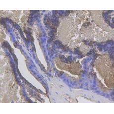
Fig3: Immunohistochemical analysis of paraffin-embedded rat skin tissue using anti-Cytokeratin 17 antibody. The section was pre-treated using heat mediated antigen retrieval with Tris-EDTA buffer (pH 8.0-8.4) for 20 minutes.The tissues were blocked in 5% BSA for 30 minutes at room temperature, washed with ddH2O and PBS, and then probed with the antibody dilution, for 30 minutes at room temperature and detected using an HRP conjugated compact polymer system. DAB was used as the chromogen. Counter stained with hematoxylin and mounted with DPX.

Fig4: Immunohistochemical analysis of paraffin-embedded human skin tissue using anti-Cytokeratin 17 antibody. The section was pre-treated using heat mediated antigen retrieval with Tris-EDTA buffer (pH 8.0-8.4) for 20 minutes.The tissues were blocked in 5% BSA for 30 minutes at room temperature, washed with ddH2O and PBS, and then probed with the antibody at 1/200 dilution, for 30 minutes at room temperature and detected using an HRP conjugated compact polymer system. DAB was used as the chromogen. Counter stained with hematoxylin and mounted with DPX.

Fig5: Immunohistochemical analysis of paraffin-embedded mouse prostate tissue using anti-Cytokeratin 17 antibody. The section was pre-treated using heat mediated antigen retrieval with Tris-EDTA buffer (pH 8.0-8.4) for 20 minutes.The tissues were blocked in 5% BSA for 30 minutes at room temperature, washed with ddH2O and PBS, and then probed with the antibody at 1/200 dilution, for 30 minutes at room temperature and detected using an HRP conjugated compact polymer system. DAB was used as the chromogen. Counter stained with hematoxylin and mounted with DPX.

Fig6: Flow cytometric analysis of Cytokeratin 17 was done on SiHa cells. The cells were fixed, permeabilized and stained with the primary antibody (red). After incubation of the primary antibody at room temperature for an hour, the cells were stained with a Alexa Fluor 488-conjugated Goat anti-Rabbit IgG Secondary antibody at 1/1000 dilution for 30 minutes.Unlabelled sample was used as a control (cells without incubation with primary antibody; black).
特别提示:本公司的所有产品仅可用于科研实验,严禁用于临床医疗及其他非科研用途!


