


-

专业包装 正品保证
-

快乐服务 售后无忧
-

会员特权 优惠不断
-

个人信息 严格保护
| 别名: | Anti-B3GAT1 antibody | ||
|---|---|---|---|
| 适用物种: | Human,Mouse,Rat | ||
| 验证应用: | WB,IHC-P,FC | ||
| 种属: | 兔多抗 | ||
| 储存条件: | -20℃ | ||
|
| 货号 | 规格 | 可用库存 | 销售价(RMB) | 您的折扣价(RMB) | 购买数量 |
|---|
| 熔点: | |
|---|---|
| 密度: | |
| 储存条件: | -20℃ |
Anti-B3GAT1 antibody
产品描述Galactosylgalactosylxylosylprotein 3-beta-glucuronosyltransferase 1 (B3GAT1) is an enzyme that in humans is encoded by the B3GAT1 gene, whose enzymatic activity creates the CD57 epitope on other cell surface proteins. In immunology, the CD57 antigen (CD stands for cluster of differentiation) is also known as HNK1 (human natural killer-1) or LEU7. It is expressed as a carbohydrate epitope that contains a sulfoglucuronyl residue in several adhesion molecules of the nervous system. The protein encoded by this gene is a member of the glucuronyltransferase gene family. These enzymes exhibit strict acceptor specificity, recognizing nonreducing terminal sugars and their anomeric linkages. This gene product functions as the key enzyme in a glucuronyl transfer reaction during the biosynthesis of the carbohydrate epitope HNK-1 (human natural killer-1, also known as CD57 and LEU7). Alternate transcriptional splice variants have been characterized. Neoplastic CD57 positive cells are seen in conditions as varied as large granular lymphocytic leukaemia, small-cell carcinoma, thyroid carcinoma, and neural and carcinoid tumours. Although the antigen is particularly common in carcinoid tumours, it is found in such a wide range of other conditions that it is of less use in distinguishing these tumours from others than more specific markers such as chromogranin and NSE.
产品名称Anti-B3GAT1 antibody
分子量38 kDa
种属反应性Human,Mouse,Rat
验证应用WB,IHC-P,FC
抗体类型兔多抗
免疫原Synthetic peptide within Human B3GAT1 aa 30-60.
偶联Non-conjugated
形态Liquid
浓度1 mg/mL.
存放说明Store at +4℃ after thawing. Aliquot store at -20℃. Avoid repeated freeze / thaw cycles.
存储缓冲液1*PBS (pH7.4), 0.2% BSA, 50% Glycerol. Preservative: 0.05% Sodium Azide.
亚型IgG
纯化方式Peptide affinity purified.
亚细胞定位Golgi apparatus. Secreted. Endoplasmic reticulum.
其它名称
WB:1:500-1:1000
IHC-P:1:50-1:200
FC:1:50-1:100

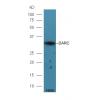
Fig1: Western blot analysis of B3GAT1 on rat brain tissue lysate. Proteins were transferred to a PVDF membrane and blocked with 5% BSA in PBS for 1 hour at room temperature. The primary antibody was used in 5% BSA at room temperature for 2 hours. Goat Anti-Rabbit IgG - HRP Secondary Antibody (HA1001) at 1:5,000 dilution was used for 1 hour at room temperature.

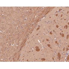
Fig2: Immunohistochemical analysis of paraffin-embedded rat brain tissue using anti-B3GAT1 antibody. The section was pre-treated using heat mediated antigen retrieval with sodium citrate buffer (pH 6.0) for 20 minutes. The tissues were blocked in 5% BSA for 30 minutes at room temperature, washed with ddH2O and PBS, and then probed with the primary antibody for 30 minutes at room temperature. The detection was performed using an HRP conjugated compact polymer system. DAB was used as the chromogen. Tissues were counterstained with hematoxylin and mounted with DPX.

Fig3: Immunohistochemical analysis of paraffin-embedded mouse brain tissue using anti-B3GAT1 antibody. The section was pre-treated using heat mediated antigen retrieval with sodium citrate buffer (pH 6.0) for 20 minutes. The tissues were blocked in 5% BSA for 30 minutes at room temperature, washed with ddH2O and PBS, and then probed with the primary antibody for 30 minutes at room temperature. The detection was performed using an HRP conjugated compact polymer system. DAB was used as the chromogen. Tissues were counterstained with hematoxylin and mounted with DPX.

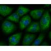
Fig4: Flow cytometric analysis of B3GAT1 was done on SH-SY5Y cells. The cells were fixed, permeabilized and stained with the primary antibody(red). After incubation of the primary antibody at room temperature for an hour, the cells were stained with a Alexa Fluor 488-conjugated Goat anti-Rabbit IgG Secondary antibody at 1/1000 dilution for 30 minutes.Unlabelled sample was used as a control (cells without incubation with primary antibody; black).
特别提示:本公司的所有产品仅可用于科研实验,严禁用于临床医疗及其他非科研用途!


