Anti-CACNA1C antibody
-
概述
- 产品描述Pore-forming, alpha-1C subunit of the voltage-gated calcium channel that gives rise to L-type calcium currents. Mediates influx of calcium ions into the cytoplasm, and thereby triggers calcium release from the sarcoplasm. Plays an important role in excitation-contraction coupling in the heart. Required for normal heart development and normal regulation of heart rhythm. Required for normal contraction of smooth muscle cells in blood vessels and in the intestine. Essential for normal blood pressure regulation via its role in the contraction of arterial smooth muscle cells. Long-lasting (L-type) calcium channels belong to the 'high-voltage activated' (HVA) group (Probable).
- 产品名称Anti-CACNA1C antibody
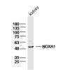
- 分子量249 kDa
- 种属反应性Human,Mouse,Rat
- 验证应用Dot blot,ICC,IHC-P,FC
- 抗体类型兔多抗
- 免疫原Synthetic peptide within human CACNA1C aa 800-880.
- 偶联Non-conjugated
-
性能
- 形态Liquid
- 浓度1 mg/mL.
- 存放说明Store at +4℃ after thawing. Aliquot store at -20℃. Avoid repeated freeze / thaw cycles.
- 存储缓冲液1*PBS (pH7.4), 0.2% BSA, 50% Glycerol. Preservative: 0.05% Sodium Azide.
- 亚型IgG
- 纯化方式Peptide affinity purified.
- 亚细胞定位Plasma membrane.
- 其它名称alpha-1 polypeptide antibody
cardiac muscle antibody
isoform 1 antibody
L type antibody
CAC1C_HUMAN antibody
CACH 2 antibody
CACH2 antibody
CACN 2 antibody
CACN2 antibody
CACNA1C antibody
CACNL1A1 antibody
Calcium channel antibody
Calcium channel cardic dihydropyridine sensitive alpha 1 subunit antibody
Calcium channel L type alpha 1 polypeptide isoform 1 cardiac muscle antibody
Calcium channel voltage dependent L type alpha 1C subunit antibody
CaV1.2 antibody
CCHL1A1 antibody
DHPR alpha 1 antibody
DHPR alpha 1 subunit antibody
LQT8 antibody
TS antibody
Voltage dependent L type calcium channel alpha 1C subunit antibody
Voltage dependent L type calcium channel subunit alpha 1C antibody
Voltage gated calcium channel alpha subunit Cav1.2 antibody
Voltage gated calcium channel subunit alpha Cav1.2 antibody
Voltage gated L type calcium channel Cav1.2 alpha 1 subunit, splice variant 10* antibody
Voltage-dependent L-type calcium channel subunit alpha-1C antibody
Voltage-gated calcium channel subunit alpha Cav1.2 antibody
more
-
应用
Dot Blot: 1:500-1:1,000
ICC: 1:500-1:2,000
IHC-P: 1:50-1:200
FC: 1:50-1:100
-
Fig1: Dot blot analysis of anti-CACNA1C on PVDF. 1ug, 2ug and 4ug of immunization peptides were given in this test. Anti-CACNA1C antibody was diluted with 1/500.
Fig2: ICC staining CACNA1C in SKOV-3 cells (green). The nuclear counter stain is DAPI (blue). Cells were fixed in paraformaldehyde, permeabilised with 0.25% Triton X100/PBS.
Fig3: Immunohistochemical analysis of paraffin-embedded rat brain tissue using anti-CACNA1C antibody. Counter stained with hematoxylin.
Fig4: Immunohistochemical analysis of paraffin-embedded human kidney tissue using anti-CACNA1C antibody. Counter stained with hematoxylin.
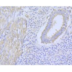
Fig5: Immunohistochemical analysis of paraffin-embedded human uterus tissue using anti-CACNA1C antibody. Counter stained with hematoxylin.
Fig6: Immunohistochemical analysis of paraffin-embedded mouse heart tissue using anti-CACNA1C antibody. Counter stained with hematoxylin.
Fig7: Flow cytometric analysis of SKOV-3 cells with CACNA1C antibody at 1/100 dilution (fuchsia) compared with an unlabelled control (cells without incubation with primary antibody; yellow). Alexa Fluor 488-conjugated goat anti-rabbit IgG was used as the
特别提示:本公司的所有产品仅可用于科研实验,严禁用于临床医疗及其他非科研用途!