Anti-MCU antibody
-
概述
- 产品描述Mitochondrial inner membrane calcium uniporter that mediates calcium uptake into mitochondria. Constitutes the pore-forming and calcium-conducting subunit of the uniporter complex. Activity is regulated by MICU1 and MICU2. At low Ca2+ levels MCU activity is down-regulated by MICU1 and MICU2; at higher Ca2+ levels MICU1 increases MCU activity. Mitochondrial calcium homeostasis plays key roles in cellular physiology and regulates cell bioenergetics, cytoplasmic calcium signals and activation of cell death pathways. Involved in buffering the amplitude of systolic calcium rises in cardiomyocytes. While dispensable for baseline homeostatic cardiac function, acts as a key regulator of short-term mitochondrial calcium loading underlying a 'fight-or-flight' response during acute stress: acts by mediating a rapid increase of mitochondrial calcium in pacemaker cells.
- 产品名称Anti-MCU antibody
- 分子量Predicted band size 40 kDa
- 种属反应性Human,Mouse,Rat
- 验证应用WB,ICC,IHC-P,FC
- 抗体类型兔多抗
- 免疫原Recombinant protein within human MCU aa 50-250.
- 偶联Non-conjugated
-
性能
- 形态Liquid
- 浓度1 mg/mL.
- 存放说明Store at +4℃ after thawing. Aliquot store at -20℃. Avoid repeated freeze / thaw cycles.
- 存储缓冲液1*PBS (pH7.4), 0.2% BSA, 50% Glycerol. Preservative: 0.05% Sodium Azide.
- 亚型IgG
- 纯化方式Protein affinity purified.
- 亚细胞定位Mitochondrion.
- 其它名称C109A_HUMAN antibody
C10orf42 antibody
Calcium uniporter protein mitochondrial antibody
Ccdc109a antibody
Coiled-coil domain-containing protein 109A antibody
HsMCU antibody
Mitochondrial Calcium Uniporter antibody
more
-
应用
WB: 1:500-1:1,000
ICC: 1:50-1:200
IHC-P: 1:50 -1:200
FC: 1:50-1:100
-
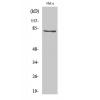
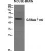
Fig1: Western blot analysis of MCU on different lysates using anti-MCU antibody at 1/500 dilution.
Positive control:
Lane 1: A549
Lane 3: mouse spleen tissue
Lane 2: HL-60
Lane 4: mouse brain tissue
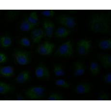
Fig2: ICC staining MCU in A431 cells (green). The nuclear counter stain is DAPI (blue). Cells were fixed in paraformaldehyde, permeabilised with 0.25% Triton X100/PBS.
Fig3: ICC staining MCU in A549 cells (green). The nuclear counter stain is DAPI (blue). Cells were fixed in paraformaldehyde, permeabilised with 0.25% Triton X100/PBS.
Fig4: ICC staining MCU in HT-29 cells (green). The nuclear counter stain is DAPI (blue). Cells were fixed in paraformaldehyde, permeabilised with 0.25% Triton X100/PBS.
Fig5: Immunohistochemical analysis of paraffin-embedded rat cerebellum tissue using anti-MCU antibody. Counter stained with hematoxylin.
Fig6: Immunohistochemical analysis of paraffin-embedded human lung cancer tissue using anti-MCU antibody. Counter stained with hematoxylin.
Fig7: Immunohistochemical analysis of paraffin-embedded mouse brain tissue using anti-MCU antibody. Counter stained with hematoxylin.
Fig8: Immunohistochemical analysis of paraffin-embedded human colon tissue using anti-MCU antibody. Counter stained with hematoxylin.
Fig9: Immunohistochemical analysis of paraffin-embedded human esophagus tissue using anti-MCU antibody. Counter stained with hematoxylin.
Fig10: Flow cytometric analysis of A549 cells with MCU antibody at 1/100 dilution (red) compared with an unlabelled control (cells without incubation with primary antibody; black). Alexa Fluor 488-conjugated goat anti-rabbit IgG was used as the secondary
特别提示:本公司的所有产品仅可用于科研实验,严禁用于临床医疗及其他非科研用途!